Doodipala Samba Reddy, Hudson Boyd, Vinitha Karnati, Joon Kim, Sreevidhya Ramakrishnan, Xin Wu
{"title":"Epigenetic Histone Deacetylases Activity Assay in the Brain and Peripheral Organ Tissues","authors":"Doodipala Samba Reddy, Hudson Boyd, Vinitha Karnati, Joon Kim, Sreevidhya Ramakrishnan, Xin Wu","doi":"10.1002/cpz1.70143","DOIUrl":null,"url":null,"abstract":"<p>Histone deacetylases (HDACs) are crucial epigenetic regulators involved in the modulation of gene expression and are promising therapeutic targets for treating various diseases, including central nervous system disorders and cancer. HDAC inhibitors exhibit neuroprotective, antiepileptogenic, and antidepressant properties in animal models, underscoring their clinical relevance. Quantifying HDAC activity is essential for identifying inhibitors and evaluating their effects under physiological or pathological conditions. This article outlines a comprehensive, sensitive, and robust assay to quantify HDAC activity in tissue lysates, with specific application to brain tissue. The assay is based on the catalytic removal of an acetyl group from the Boc-Lys(Ac)-AMC substrate by HDAC enzymes. Following nuclear protein extraction, tissue HDAC activity can be quantitatively assessed using a fluorometric Boc-Lys(Ac) HDAC activity kit. Adding a developer containing trypsin then converts the deacetylated product into a measurable fluorophore. This enables precise quantification of HDAC activity levels across different tissues, making the method suitable for screening putative HDAC inhibitors and assessing their effects on epigenetically modulated phenotypes. We validated these protocols using brain tissue samples from mice subjected to traumatic brain injury, a condition known to elevate HDAC activity levels. This assay provides an efficient, scalable tool for exploring HDAC function, evaluating therapeutic interventions, and advancing the understanding of HDAC-mediated mechanisms in animal models. © 2025 The Author(s). Current Protocols published by Wiley Periodicals LLC.</p><p><b>Basic Protocol 1</b>: Isolation of nuclear protein from brain and other tissues</p><p><b>Support Protocol 1</b>: Harvesting and microdissection of brain and other tissues</p><p><b>Support Protocol 2</b>: Estimation of extracted protein using the Pierce bicinchoninic acid (BCA) assay</p><p><b>Basic Protocol 2</b>: HDAC activity fluorometric assay in the brain and other tissues</p>","PeriodicalId":93970,"journal":{"name":"Current protocols","volume":"5 6","pages":""},"PeriodicalIF":2.2000,"publicationDate":"2025-06-11","publicationTypes":"Journal Article","fieldsOfStudy":null,"isOpenAccess":false,"openAccessPdf":"https://onlinelibrary.wiley.com/doi/epdf/10.1002/cpz1.70143","citationCount":"0","resultStr":null,"platform":"Semanticscholar","paperid":null,"PeriodicalName":"Current protocols","FirstCategoryId":"1085","ListUrlMain":"https://currentprotocols.onlinelibrary.wiley.com/doi/10.1002/cpz1.70143","RegionNum":0,"RegionCategory":null,"ArticlePicture":[],"TitleCN":null,"AbstractTextCN":null,"PMCID":null,"EPubDate":"","PubModel":"","JCR":"","JCRName":"","Score":null,"Total":0}
引用次数: 0
Abstract
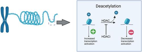
Histone deacetylases (HDACs) are crucial epigenetic regulators involved in the modulation of gene expression and are promising therapeutic targets for treating various diseases, including central nervous system disorders and cancer. HDAC inhibitors exhibit neuroprotective, antiepileptogenic, and antidepressant properties in animal models, underscoring their clinical relevance. Quantifying HDAC activity is essential for identifying inhibitors and evaluating their effects under physiological or pathological conditions. This article outlines a comprehensive, sensitive, and robust assay to quantify HDAC activity in tissue lysates, with specific application to brain tissue. The assay is based on the catalytic removal of an acetyl group from the Boc-Lys(Ac)-AMC substrate by HDAC enzymes. Following nuclear protein extraction, tissue HDAC activity can be quantitatively assessed using a fluorometric Boc-Lys(Ac) HDAC activity kit. Adding a developer containing trypsin then converts the deacetylated product into a measurable fluorophore. This enables precise quantification of HDAC activity levels across different tissues, making the method suitable for screening putative HDAC inhibitors and assessing their effects on epigenetically modulated phenotypes. We validated these protocols using brain tissue samples from mice subjected to traumatic brain injury, a condition known to elevate HDAC activity levels. This assay provides an efficient, scalable tool for exploring HDAC function, evaluating therapeutic interventions, and advancing the understanding of HDAC-mediated mechanisms in animal models. © 2025 The Author(s). Current Protocols published by Wiley Periodicals LLC.
Basic Protocol 1: Isolation of nuclear protein from brain and other tissues
Support Protocol 1: Harvesting and microdissection of brain and other tissues
Support Protocol 2: Estimation of extracted protein using the Pierce bicinchoninic acid (BCA) assay
Basic Protocol 2: HDAC activity fluorometric assay in the brain and other tissues


 求助内容:
求助内容: 应助结果提醒方式:


