Sunil Buddappa, Rajesh Kumar Barooah, B K Baishya, Fazlallah Afshangian, Daniel Encarnacion-Santos, Gianluca Scalia, Giuseppe E Umana, Bipin Chaurasia
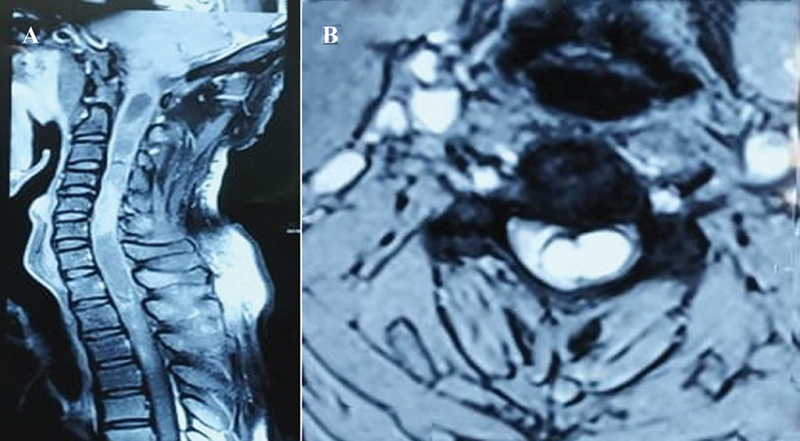
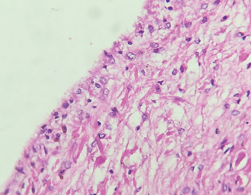
{"title":"Isolated Intramedullary Spinal Neurenteric Cysts: Case Report and Literature Review.","authors":"Sunil Buddappa, Rajesh Kumar Barooah, B K Baishya, Fazlallah Afshangian, Daniel Encarnacion-Santos, Gianluca Scalia, Giuseppe E Umana, Bipin Chaurasia","doi":"10.1055/s-0045-1806727","DOIUrl":null,"url":null,"abstract":"<p><p>Neurenteric cysts, also known as endodermal or enterogenous cysts, are uncommon benign congenital lesions of the central nervous system (CNS) characterized by an epithelial lining of endodermal origin. These cysts predominantly affect the spinal canal and cord. Intramedullary neurenteric cysts are exceptionally rare, with fewer than 100 reported isolated cases. Their distinct characteristics, clinical presentation, and challenges in diagnosis and treatment necessitate a detailed case analysis and review. We present the case of a 33-year-old male patient with an intriguing case of an isolated intramedullary cystic lesion in the cervicodorsal spinal cord, extending from the cervicobulbar junction to the D4 vertebra level. The patient's clinical presentation included a 6-month history of progressive weakness in the left upper limb, accompanied by pain and numbness. Neurological examination revealed muscle atrophy, reduced strength, spastic paraparesis, and sensory deficits. Radiological findings demonstrated an expansile cystic lesion with marked signal heterogeneity, intense enhancement, and the presence of a \"cap sign\" indicative of subacute hemorrhage. Diagnosis of spinal intramedullary neurenteric cysts is reliant on histopathology. Surgical removal remains the recommended treatment, as a conservative approach can lead to irreversible neurological deficits. However, complete resection may be challenging due to potential adhesions to surrounding structures. In such cases, a more conservative approach, avoiding cyst spillage into the subarachnoid space, is preferred. Vigilant radiological follow-up is crucial to monitor for potential cyst recurrence. These rare cases highlight the need for further scientific literature and improved diagnostic and therapeutic strategies.</p>","PeriodicalId":94300,"journal":{"name":"Asian journal of neurosurgery","volume":"20 2","pages":"408-412"},"PeriodicalIF":0.0000,"publicationDate":"2025-03-18","publicationTypes":"Journal Article","fieldsOfStudy":null,"isOpenAccess":false,"openAccessPdf":"https://www.ncbi.nlm.nih.gov/pmc/articles/PMC12136934/pdf/","citationCount":"0","resultStr":null,"platform":"Semanticscholar","paperid":null,"PeriodicalName":"Asian journal of neurosurgery","FirstCategoryId":"1085","ListUrlMain":"https://doi.org/10.1055/s-0045-1806727","RegionNum":0,"RegionCategory":null,"ArticlePicture":[],"TitleCN":null,"AbstractTextCN":null,"PMCID":null,"EPubDate":"2025/6/1 0:00:00","PubModel":"eCollection","JCR":"","JCRName":"","Score":null,"Total":0}
引用次数: 0
Abstract
Neurenteric cysts, also known as endodermal or enterogenous cysts, are uncommon benign congenital lesions of the central nervous system (CNS) characterized by an epithelial lining of endodermal origin. These cysts predominantly affect the spinal canal and cord. Intramedullary neurenteric cysts are exceptionally rare, with fewer than 100 reported isolated cases. Their distinct characteristics, clinical presentation, and challenges in diagnosis and treatment necessitate a detailed case analysis and review. We present the case of a 33-year-old male patient with an intriguing case of an isolated intramedullary cystic lesion in the cervicodorsal spinal cord, extending from the cervicobulbar junction to the D4 vertebra level. The patient's clinical presentation included a 6-month history of progressive weakness in the left upper limb, accompanied by pain and numbness. Neurological examination revealed muscle atrophy, reduced strength, spastic paraparesis, and sensory deficits. Radiological findings demonstrated an expansile cystic lesion with marked signal heterogeneity, intense enhancement, and the presence of a "cap sign" indicative of subacute hemorrhage. Diagnosis of spinal intramedullary neurenteric cysts is reliant on histopathology. Surgical removal remains the recommended treatment, as a conservative approach can lead to irreversible neurological deficits. However, complete resection may be challenging due to potential adhesions to surrounding structures. In such cases, a more conservative approach, avoiding cyst spillage into the subarachnoid space, is preferred. Vigilant radiological follow-up is crucial to monitor for potential cyst recurrence. These rare cases highlight the need for further scientific literature and improved diagnostic and therapeutic strategies.

 求助内容:
求助内容: 应助结果提醒方式:


