Cerebellar Mutism/Posterior Fossa Syndrome Following Resection of Posterior Fossa Tumor in Pediatric Patients: Assessing Pathophysiology, Risk Factors, and Neuroradiographic Features.
{"title":"Cerebellar Mutism/Posterior Fossa Syndrome Following Resection of Posterior Fossa Tumor in Pediatric Patients: Assessing Pathophysiology, Risk Factors, and Neuroradiographic Features.","authors":"Vikrant Setia, Monirah Zeya, Arvind Kumar Srivastava, Anita Jagetia","doi":"10.1055/s-0044-1801404","DOIUrl":null,"url":null,"abstract":"<p><strong>Background: </strong>Cerebellar mutism syndrome (CMS) is a postoperative syndrome of decreased speech seen in children associated with neurobehavioral abnormalities, the incidence of which is up to 40%.</p><p><strong>Objectives: </strong>To evaluate pediatric patients with posterior fossa tumors for incidence, clinical characteristics, pathophysiology, risk factors, and neuroradiographic features of this syndrome.</p><p><strong>Materials and methods: </strong>The study included 60 pediatric patients with a posterior fossa tumor who underwent surgery by a telovelar approach. Detailed pre- and postoperative clinical and radiological evaluations were done. Patients with CMS were analyzed and compared with those without mutism to find risk factors for CMS. The presentation and characteristics of cerebellar mutism were studied along with the following risk factors:Clinical-age, sex, cranial nerve deficit, and adjuvant treatment.Radiological-tumor location, hydrocephalus, brainstem invasion, extent of tumor resection, peduncular and brainstem edema, and atrophy of posterior fossa structures.Pathological-histopathology of tumor.The preoperative, immediate postoperative, and 1-year postoperative imaging results were reviewed to assess the neuroradiographic features in the two groups.</p><p><strong>Results: </strong>The incidence of this syndrome was 20%. The mutism was accompanied by some neurobehavioral abnormalities ( <i>p</i> -value = 0.05). The most significant finding was the presence of a period of cerebellar dysarthria after the resolution of the muteness ( <i>p</i> -value < 0.001) in all cases. Brainstem and related structures' involvement was the most significant risk factor ( <i>p</i> -value = 0.03). The presence of brainstem and peduncular edema in the immediate postoperative period ( <i>p</i> -value = 0.04) and gross atrophy of posterior fossa structures at 1 year ( <i>p</i> -value = 0.01) showed significance toward the development of CMS. There was delayed neurological recovery in patients with CMS with a poor Glasgow Outcome Score at 1 year of follow-up.</p><p><strong>Conclusion: </strong>The clinical presentation of this syndrome in context with neuroradiographic features suggests that it results from transient impairment of the afferent and/or efferent pathways of dentate nuclei that are involved in initiating complex volitional movements and are associated with brainstem involvement of tumor and poor functional outcome.</p>","PeriodicalId":94300,"journal":{"name":"Asian journal of neurosurgery","volume":"20 2","pages":"260-268"},"PeriodicalIF":0.0000,"publicationDate":"2025-01-13","publicationTypes":"Journal Article","fieldsOfStudy":null,"isOpenAccess":false,"openAccessPdf":"https://www.ncbi.nlm.nih.gov/pmc/articles/PMC12136955/pdf/","citationCount":"0","resultStr":null,"platform":"Semanticscholar","paperid":null,"PeriodicalName":"Asian journal of neurosurgery","FirstCategoryId":"1085","ListUrlMain":"https://doi.org/10.1055/s-0044-1801404","RegionNum":0,"RegionCategory":null,"ArticlePicture":[],"TitleCN":null,"AbstractTextCN":null,"PMCID":null,"EPubDate":"2025/6/1 0:00:00","PubModel":"eCollection","JCR":"","JCRName":"","Score":null,"Total":0}
引用次数: 0
Abstract
Background: Cerebellar mutism syndrome (CMS) is a postoperative syndrome of decreased speech seen in children associated with neurobehavioral abnormalities, the incidence of which is up to 40%.
Objectives: To evaluate pediatric patients with posterior fossa tumors for incidence, clinical characteristics, pathophysiology, risk factors, and neuroradiographic features of this syndrome.
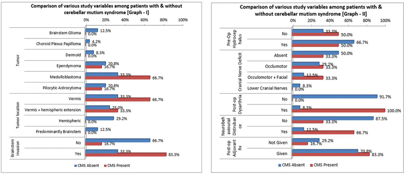
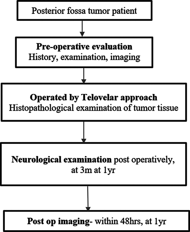
Materials and methods: The study included 60 pediatric patients with a posterior fossa tumor who underwent surgery by a telovelar approach. Detailed pre- and postoperative clinical and radiological evaluations were done. Patients with CMS were analyzed and compared with those without mutism to find risk factors for CMS. The presentation and characteristics of cerebellar mutism were studied along with the following risk factors:Clinical-age, sex, cranial nerve deficit, and adjuvant treatment.Radiological-tumor location, hydrocephalus, brainstem invasion, extent of tumor resection, peduncular and brainstem edema, and atrophy of posterior fossa structures.Pathological-histopathology of tumor.The preoperative, immediate postoperative, and 1-year postoperative imaging results were reviewed to assess the neuroradiographic features in the two groups.
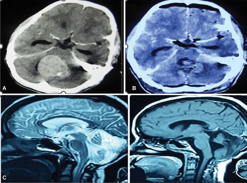
Results: The incidence of this syndrome was 20%. The mutism was accompanied by some neurobehavioral abnormalities ( p -value = 0.05). The most significant finding was the presence of a period of cerebellar dysarthria after the resolution of the muteness ( p -value < 0.001) in all cases. Brainstem and related structures' involvement was the most significant risk factor ( p -value = 0.03). The presence of brainstem and peduncular edema in the immediate postoperative period ( p -value = 0.04) and gross atrophy of posterior fossa structures at 1 year ( p -value = 0.01) showed significance toward the development of CMS. There was delayed neurological recovery in patients with CMS with a poor Glasgow Outcome Score at 1 year of follow-up.
Conclusion: The clinical presentation of this syndrome in context with neuroradiographic features suggests that it results from transient impairment of the afferent and/or efferent pathways of dentate nuclei that are involved in initiating complex volitional movements and are associated with brainstem involvement of tumor and poor functional outcome.

 求助内容:
求助内容: 应助结果提醒方式:


