Anis Choucha, Matteo De Simone, Nathan Beucler, Solenne Hulot, Jean-Christophe Lagier, Henry Dufour
{"title":"Brain Abscess Mimicking Brain Tumors: A Systematic Review of Individual Patient's Data.","authors":"Anis Choucha, Matteo De Simone, Nathan Beucler, Solenne Hulot, Jean-Christophe Lagier, Henry Dufour","doi":"10.1055/s-0045-1802623","DOIUrl":null,"url":null,"abstract":"<p><p><b>Objectives</b> Brain abscess is a worrisome condition with a 1-year mortality rate of 21% and a 32% rate of new-onset epilepsy. Brain magnetic resonance imaging (MRI) is strongly recommended as a screening modality with contrast-enhanced T1-weighted images, diffusion-weighted imaging (DWI), and attenuated diffusion coefficient. However, there is a 10% rate of false negative, which could potentially impact management and prognosis. Our systematic review aims at identifying risk factors for false negative. <b>Materials and Methods</b> A database search of our institutions plus a systematic literature review was conducted using MEDLINE/PubMed, including studies of brain abscesses misdiagnosed as brain tumors. Data on patient demographics, clinical presentations, imaging findings, pathogens, treatments, and outcomes were extracted and analyzed. We present a case of a 59-year-old male with HIV, who developed a brain abscess misdiagnosed as a tumor. Initial symptoms included left-side weakness and weight loss. Imaging showed a ring-enhancing lesion in the right thalamus. The abscess was caused by <i>T. gondii</i> , and the patient was treated with sulfadiazine, pyrimethamine, ceftriaxone, and metronidazole, achieving a GOS-E score of 8 at 1 year. <b>Results</b> The review included 14 studies, with 1 additional illustrative case, encompassing a total of 15 cases. Patients ranged from 39 to 77 years, with a mean age of 59 years. Comorbidities included human immunodeficiency virus (HIV), glioblastoma, breast cancer, arthritis, gastric cancer, and nephrotic syndrome. Common symptoms were hemiparesis, generalized seizures, headache, and confusion. Imaging often revealed ring-enhancing lesions with restricted diffusion on DWI. Lesions were located in various brain regions. Pathogens identified included 40% <i>Nocardia</i> species, <i>Toxoplasma gondii</i> , <i>Mycobacterium tuberculosis</i> , <i>Aggregatibacter aphrophilus</i> , <i>Rickettsia typhi</i> , <i>Arcanobacterium haemolyticum</i> , <i>Aspergillus terreus</i> , and <i>Providencia rettgeri</i> . Treatments involved antibiotics and, in some cases, surgical intervention. Outcomes measured by the Glasgow Outcome Scale-Extended (GOS-E) at 1 year indicated good recovery in most cases. <b>Conclusion</b> Despite the high sensitivity and specificity of brain MRI in diagnosing brain abscesses, the standard protocol used for the past two decades still results in a 10% false-negative rate. Such inaccuracies can significantly impact the patient's management, potentially delaying antibiotic therapy and impacting the surgical planning, hence affecting the outcome. Immunocompromised patients are particularly vulnerable to misdiagnoses of brain abscesses as brain tumors. To improve diagnostic accuracy, new imaging techniques and computational tools are currently under investigation.</p>","PeriodicalId":94300,"journal":{"name":"Asian journal of neurosurgery","volume":"20 2","pages":"291-300"},"PeriodicalIF":0.0000,"publicationDate":"2025-02-06","publicationTypes":"Journal Article","fieldsOfStudy":null,"isOpenAccess":false,"openAccessPdf":"https://www.ncbi.nlm.nih.gov/pmc/articles/PMC12136936/pdf/","citationCount":"0","resultStr":null,"platform":"Semanticscholar","paperid":null,"PeriodicalName":"Asian journal of neurosurgery","FirstCategoryId":"1085","ListUrlMain":"https://doi.org/10.1055/s-0045-1802623","RegionNum":0,"RegionCategory":null,"ArticlePicture":[],"TitleCN":null,"AbstractTextCN":null,"PMCID":null,"EPubDate":"2025/6/1 0:00:00","PubModel":"eCollection","JCR":"","JCRName":"","Score":null,"Total":0}
引用次数: 0
Abstract
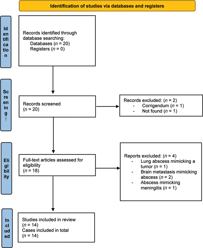
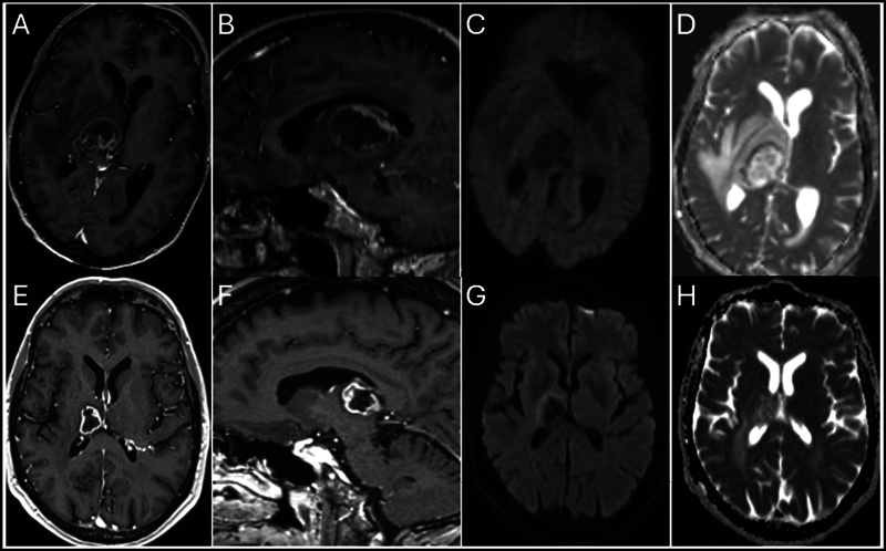
Objectives Brain abscess is a worrisome condition with a 1-year mortality rate of 21% and a 32% rate of new-onset epilepsy. Brain magnetic resonance imaging (MRI) is strongly recommended as a screening modality with contrast-enhanced T1-weighted images, diffusion-weighted imaging (DWI), and attenuated diffusion coefficient. However, there is a 10% rate of false negative, which could potentially impact management and prognosis. Our systematic review aims at identifying risk factors for false negative. Materials and Methods A database search of our institutions plus a systematic literature review was conducted using MEDLINE/PubMed, including studies of brain abscesses misdiagnosed as brain tumors. Data on patient demographics, clinical presentations, imaging findings, pathogens, treatments, and outcomes were extracted and analyzed. We present a case of a 59-year-old male with HIV, who developed a brain abscess misdiagnosed as a tumor. Initial symptoms included left-side weakness and weight loss. Imaging showed a ring-enhancing lesion in the right thalamus. The abscess was caused by T. gondii , and the patient was treated with sulfadiazine, pyrimethamine, ceftriaxone, and metronidazole, achieving a GOS-E score of 8 at 1 year. Results The review included 14 studies, with 1 additional illustrative case, encompassing a total of 15 cases. Patients ranged from 39 to 77 years, with a mean age of 59 years. Comorbidities included human immunodeficiency virus (HIV), glioblastoma, breast cancer, arthritis, gastric cancer, and nephrotic syndrome. Common symptoms were hemiparesis, generalized seizures, headache, and confusion. Imaging often revealed ring-enhancing lesions with restricted diffusion on DWI. Lesions were located in various brain regions. Pathogens identified included 40% Nocardia species, Toxoplasma gondii , Mycobacterium tuberculosis , Aggregatibacter aphrophilus , Rickettsia typhi , Arcanobacterium haemolyticum , Aspergillus terreus , and Providencia rettgeri . Treatments involved antibiotics and, in some cases, surgical intervention. Outcomes measured by the Glasgow Outcome Scale-Extended (GOS-E) at 1 year indicated good recovery in most cases. Conclusion Despite the high sensitivity and specificity of brain MRI in diagnosing brain abscesses, the standard protocol used for the past two decades still results in a 10% false-negative rate. Such inaccuracies can significantly impact the patient's management, potentially delaying antibiotic therapy and impacting the surgical planning, hence affecting the outcome. Immunocompromised patients are particularly vulnerable to misdiagnoses of brain abscesses as brain tumors. To improve diagnostic accuracy, new imaging techniques and computational tools are currently under investigation.

 求助内容:
求助内容: 应助结果提醒方式:


