{"title":"Anterior Clinoid Process Metastasis with Sudden Loss of Vision: Role of Emergency Optic Nerve Decompression.","authors":"Marta Rico Pereira, Fernando Muñoz Hernández","doi":"10.1055/s-0045-1806729","DOIUrl":null,"url":null,"abstract":"<p><p>Anterior clinoid process metastases are rare. We present an unusual case of anterior clinoid process metastasis with sudden deterioration of visual function requiring emergency optic nerve decompression, resulting in recovery of visual function. The patient was a 41-year-old man with a diagnosis of leiomyosarcoma of the radius, operated on in 2014, with bone and lung metastases, who had been treated with chemotherapy and appeared to have stable disease at his last follow-up. Six years later, he developed a 1-month history of progressive unilateral loss of visual acuity and visual field defect (initially quadrantanopia that progressed to nasal hemianopia). Brain imaging showed a contrast-enhancing lesion affecting the left anterior clinoid process with extension to the cavernous sinus and sphenoid sinus, causing compression of the left optic nerve. Although the lesion could have suggested a meningioma given the location, in the context of the patient's oncological history, the diagnosis of metastasis was considered more likely. The patient was admitted to the hospital and, during the hospital stay, developed sudden left retro-orbital pain progressing to left amaurosis over approximately 8 hours. Urgent surgery was performed: a pterional craniotomy with partial tumor removal and optic nerve decompression with extradural anterior clinoidectomy. After surgery, the patient had an immediate but partial improvement in visual acuity and in the visual field defect. Metastasis to the anterior clinoid process is very uncommon, with only one case previously reported in the literature. In cases of visual impairment, symptoms may deteriorate rapidly to complete loss of vision, so urgent decompressive surgery of the optic pathway may be indicated to recover visual function, although recovery may be partial.</p>","PeriodicalId":94300,"journal":{"name":"Asian journal of neurosurgery","volume":"20 2","pages":"423-426"},"PeriodicalIF":0.0000,"publicationDate":"2025-03-24","publicationTypes":"Journal Article","fieldsOfStudy":null,"isOpenAccess":false,"openAccessPdf":"https://www.ncbi.nlm.nih.gov/pmc/articles/PMC12136971/pdf/","citationCount":"0","resultStr":null,"platform":"Semanticscholar","paperid":null,"PeriodicalName":"Asian journal of neurosurgery","FirstCategoryId":"1085","ListUrlMain":"https://doi.org/10.1055/s-0045-1806729","RegionNum":0,"RegionCategory":null,"ArticlePicture":[],"TitleCN":null,"AbstractTextCN":null,"PMCID":null,"EPubDate":"2025/6/1 0:00:00","PubModel":"eCollection","JCR":"","JCRName":"","Score":null,"Total":0}
引用次数: 0
Abstract
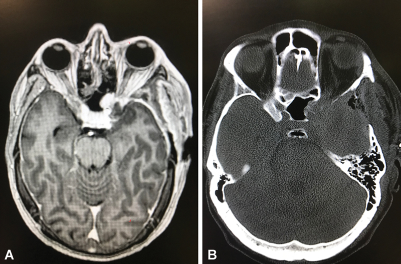
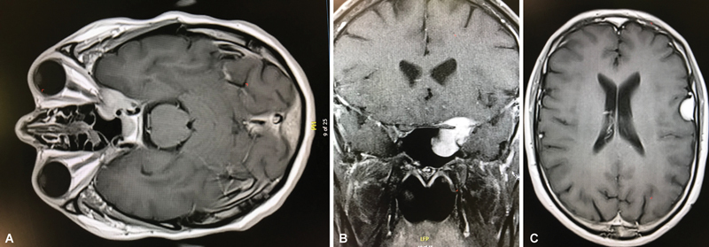
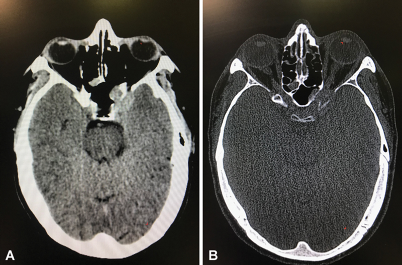
Anterior clinoid process metastases are rare. We present an unusual case of anterior clinoid process metastasis with sudden deterioration of visual function requiring emergency optic nerve decompression, resulting in recovery of visual function. The patient was a 41-year-old man with a diagnosis of leiomyosarcoma of the radius, operated on in 2014, with bone and lung metastases, who had been treated with chemotherapy and appeared to have stable disease at his last follow-up. Six years later, he developed a 1-month history of progressive unilateral loss of visual acuity and visual field defect (initially quadrantanopia that progressed to nasal hemianopia). Brain imaging showed a contrast-enhancing lesion affecting the left anterior clinoid process with extension to the cavernous sinus and sphenoid sinus, causing compression of the left optic nerve. Although the lesion could have suggested a meningioma given the location, in the context of the patient's oncological history, the diagnosis of metastasis was considered more likely. The patient was admitted to the hospital and, during the hospital stay, developed sudden left retro-orbital pain progressing to left amaurosis over approximately 8 hours. Urgent surgery was performed: a pterional craniotomy with partial tumor removal and optic nerve decompression with extradural anterior clinoidectomy. After surgery, the patient had an immediate but partial improvement in visual acuity and in the visual field defect. Metastasis to the anterior clinoid process is very uncommon, with only one case previously reported in the literature. In cases of visual impairment, symptoms may deteriorate rapidly to complete loss of vision, so urgent decompressive surgery of the optic pathway may be indicated to recover visual function, although recovery may be partial.

 求助内容:
求助内容: 应助结果提醒方式:


