Nur Liyana Mohammed Yusof, Derek M Yellon, Sean M Davidson
{"title":"The contribution of cardiomyocyte hypercontracture to the burden of acute myocardial infarction: an update.","authors":"Nur Liyana Mohammed Yusof, Derek M Yellon, Sean M Davidson","doi":"10.1007/s00395-025-01120-1","DOIUrl":null,"url":null,"abstract":"<p><p>Although reperfusion therapy such as percutaneous coronary intervention and thrombolysis have been implemented in clinical practise as treatments for acute myocardial infarction (AMI) since the 1970s, patients continue to experience high rates of morbidity and mortality. Coronary reperfusion is effective as it limits infarction. However, it induces significant myocardial injury, known as ischaemia-reperfusion (IR) injury. Sustained depletion of cellular adenosine triphosphate (ATP) leading to intracellular calcium (Ca<sup>2+</sup>) overload ultimately lead to cardiomyocyte death during ischaemia. Reperfusion enables resynthesis of ATP, but if this occurs whilst Ca<sup>2+</sup> remains elevated, it induces excessive cardiomyocyte contracture, known as hypercontracture. Irreversible myocardial injury caused by hypercontracture is often accompanied by histological findings such as wavy myocardial fibres, and more profoundly, contraction band necrosis, identified by the presence of dense eosinophilic bands within the cardiomyocytes. The presence of hypercontracture imposes deleterious effects on both cardiac function and clinical outcomes in individuals experiencing AMI. The potential cardioprotective benefits of inhibiting hypercontracture following IR injury have been demonstrated in animal models, however therapies suitable for clinical application are yet to be developed. This article reviews the pathogenesis and clinical manifestation of hypercontracture in cardiomyocytes during AMI. In addition, the discussion highlights the challenges of translating robust pre-clinical data into successful clinical therapeutic approaches.</p>","PeriodicalId":8723,"journal":{"name":"Basic Research in Cardiology","volume":" ","pages":"619-639"},"PeriodicalIF":8.0000,"publicationDate":"2025-08-01","publicationTypes":"Journal Article","fieldsOfStudy":null,"isOpenAccess":false,"openAccessPdf":"https://www.ncbi.nlm.nih.gov/pmc/articles/PMC12325465/pdf/","citationCount":"0","resultStr":null,"platform":"Semanticscholar","paperid":null,"PeriodicalName":"Basic Research in Cardiology","FirstCategoryId":"3","ListUrlMain":"https://doi.org/10.1007/s00395-025-01120-1","RegionNum":1,"RegionCategory":"医学","ArticlePicture":[],"TitleCN":null,"AbstractTextCN":null,"PMCID":null,"EPubDate":"2025/6/7 0:00:00","PubModel":"Epub","JCR":"Q1","JCRName":"CARDIAC & CARDIOVASCULAR SYSTEMS","Score":null,"Total":0}
引用次数: 0
Abstract
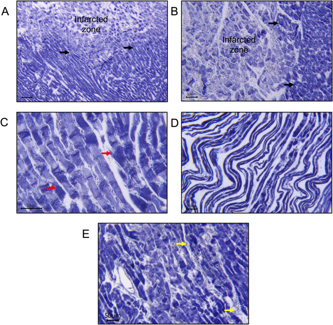
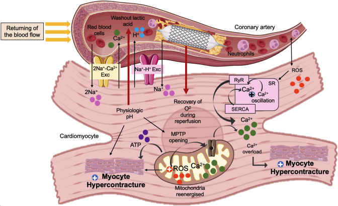
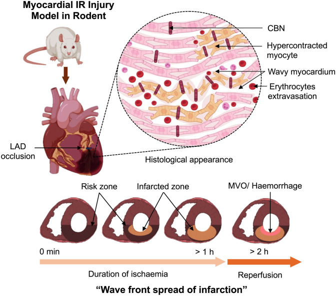
Although reperfusion therapy such as percutaneous coronary intervention and thrombolysis have been implemented in clinical practise as treatments for acute myocardial infarction (AMI) since the 1970s, patients continue to experience high rates of morbidity and mortality. Coronary reperfusion is effective as it limits infarction. However, it induces significant myocardial injury, known as ischaemia-reperfusion (IR) injury. Sustained depletion of cellular adenosine triphosphate (ATP) leading to intracellular calcium (Ca2+) overload ultimately lead to cardiomyocyte death during ischaemia. Reperfusion enables resynthesis of ATP, but if this occurs whilst Ca2+ remains elevated, it induces excessive cardiomyocyte contracture, known as hypercontracture. Irreversible myocardial injury caused by hypercontracture is often accompanied by histological findings such as wavy myocardial fibres, and more profoundly, contraction band necrosis, identified by the presence of dense eosinophilic bands within the cardiomyocytes. The presence of hypercontracture imposes deleterious effects on both cardiac function and clinical outcomes in individuals experiencing AMI. The potential cardioprotective benefits of inhibiting hypercontracture following IR injury have been demonstrated in animal models, however therapies suitable for clinical application are yet to be developed. This article reviews the pathogenesis and clinical manifestation of hypercontracture in cardiomyocytes during AMI. In addition, the discussion highlights the challenges of translating robust pre-clinical data into successful clinical therapeutic approaches.
期刊介绍:
Basic Research in Cardiology is an international journal for cardiovascular research. It provides a forum for original and review articles related to experimental cardiology that meet its stringent scientific standards.
Basic Research in Cardiology regularly receives articles from the fields of
- Molecular and Cellular Biology
- Biochemistry
- Biophysics
- Pharmacology
- Physiology and Pathology
- Clinical Cardiology


 求助内容:
求助内容: 应助结果提醒方式:


