Ümmügülsüm Yıldız-Altay, Laura J Burns, Li-Chi Chen, Himanee Parag Dave, Mariko R Yasuda, Jillian M Richmond, Maryanne M Senna
{"title":"M2 Macrophage and Extracellular Matrix Genes Are Enriched in High-Activity Lichen Planopilaris.","authors":"Ümmügülsüm Yıldız-Altay, Laura J Burns, Li-Chi Chen, Himanee Parag Dave, Mariko R Yasuda, Jillian M Richmond, Maryanne M Senna","doi":"10.1155/drp/5545886","DOIUrl":null,"url":null,"abstract":"<p><p>The pathophysiology of lichen planopilaris (LPP), a lymphocytic primary cicatricial alopecia, is largely unknown. We evaluated RNA expression of lesional scalp biopsies taken before and after 6 months of treatment monotherapy with oral hydroxychloroquine (HCQ), narrow band ultraviolet B (NB-UVB), or low level laser light therapy (LLLLT). <i>PTGER4</i> and <i>DOCK2</i> were significantly increased in all patients after treatment. <i>CYP1A2</i>, a drug metabolism enzyme, and <i>SSR2</i>, a gene involved in B cell activation and maturation, were increased posttreatment for the HCQ arm. <i>VEGFA</i>, which has been reported to be downregulated by phototherapy was decreased post NB-UVB treatment, while <i>SAA1</i>, an apolipoprotein gene present in plasma that is upregulated in response to tissue injury, was increased posttreatment for the NB-UVB arm. No significant differentially expressed genes (DEGs) in the LLLLT arm before and after treatment. The expressions of <i>CD68</i>, <i>COL5A1</i>, <i>MMP9</i>, <i>COL6A3</i>, and <i>CD44</i> were significantly higher at the baseline in biopsies from patients with a Lichen Planopilaris Activity Index (LPPAI) score ≥ 4 compared with those with an LPPAI < 4. These genes are involved in extracellular matrix organization and M2, or profibrotic, macrophage polarization, which is congruent with follicular scarring. Our data identify potential RNA biomarkers of LPPAI and suggest that M2 macrophages may play a role in LPP immunopathogenesis.</p>","PeriodicalId":11338,"journal":{"name":"Dermatology Research and Practice","volume":"2025 ","pages":"5545886"},"PeriodicalIF":1.9000,"publicationDate":"2025-05-29","publicationTypes":"Journal Article","fieldsOfStudy":null,"isOpenAccess":false,"openAccessPdf":"https://www.ncbi.nlm.nih.gov/pmc/articles/PMC12140823/pdf/","citationCount":"0","resultStr":null,"platform":"Semanticscholar","paperid":null,"PeriodicalName":"Dermatology Research and Practice","FirstCategoryId":"1085","ListUrlMain":"https://doi.org/10.1155/drp/5545886","RegionNum":0,"RegionCategory":null,"ArticlePicture":[],"TitleCN":null,"AbstractTextCN":null,"PMCID":null,"EPubDate":"2025/1/1 0:00:00","PubModel":"eCollection","JCR":"Q3","JCRName":"DERMATOLOGY","Score":null,"Total":0}
引用次数: 0
Abstract
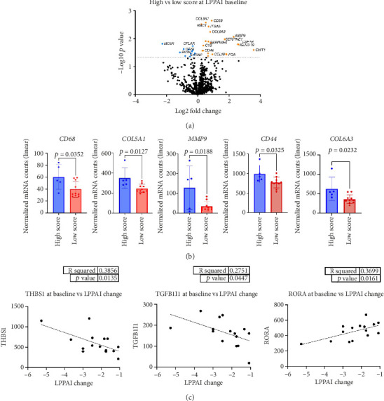
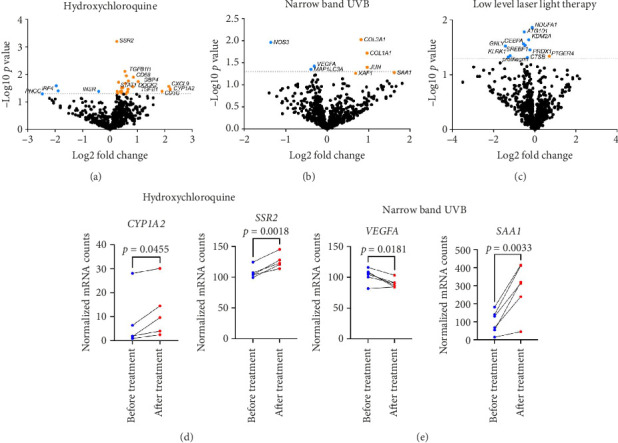
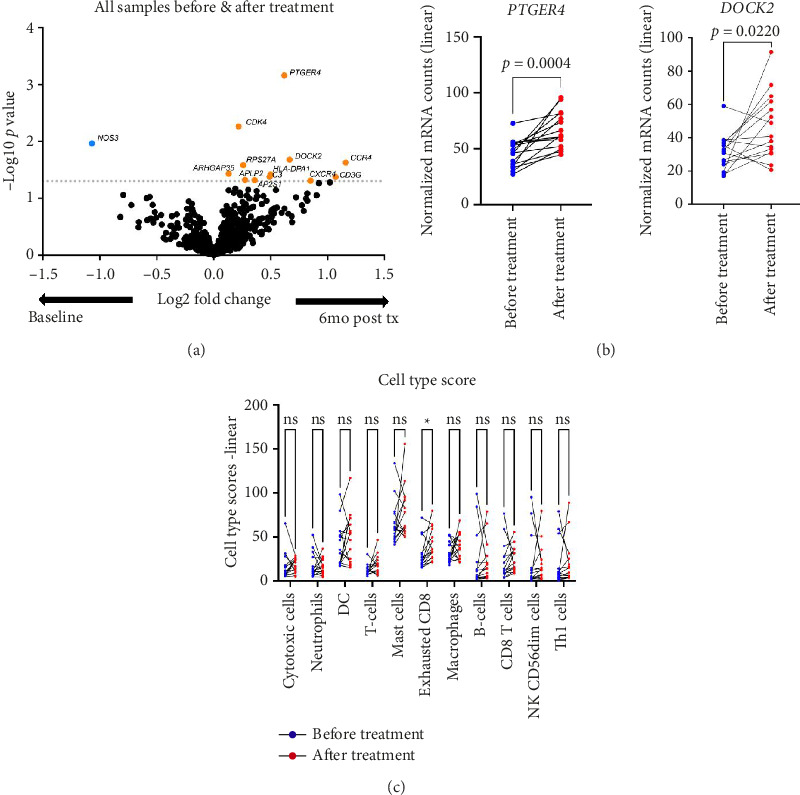
The pathophysiology of lichen planopilaris (LPP), a lymphocytic primary cicatricial alopecia, is largely unknown. We evaluated RNA expression of lesional scalp biopsies taken before and after 6 months of treatment monotherapy with oral hydroxychloroquine (HCQ), narrow band ultraviolet B (NB-UVB), or low level laser light therapy (LLLLT). PTGER4 and DOCK2 were significantly increased in all patients after treatment. CYP1A2, a drug metabolism enzyme, and SSR2, a gene involved in B cell activation and maturation, were increased posttreatment for the HCQ arm. VEGFA, which has been reported to be downregulated by phototherapy was decreased post NB-UVB treatment, while SAA1, an apolipoprotein gene present in plasma that is upregulated in response to tissue injury, was increased posttreatment for the NB-UVB arm. No significant differentially expressed genes (DEGs) in the LLLLT arm before and after treatment. The expressions of CD68, COL5A1, MMP9, COL6A3, and CD44 were significantly higher at the baseline in biopsies from patients with a Lichen Planopilaris Activity Index (LPPAI) score ≥ 4 compared with those with an LPPAI < 4. These genes are involved in extracellular matrix organization and M2, or profibrotic, macrophage polarization, which is congruent with follicular scarring. Our data identify potential RNA biomarkers of LPPAI and suggest that M2 macrophages may play a role in LPP immunopathogenesis.

 求助内容:
求助内容: 应助结果提醒方式:


