Musawenkosi M Mthombeni, Nasreen Mahomed, Grace Rubin, Sharadini K Gounden
{"title":"A review of papillary breast carcinoma in women attending a breast imaging centre in Johannesburg.","authors":"Musawenkosi M Mthombeni, Nasreen Mahomed, Grace Rubin, Sharadini K Gounden","doi":"10.4102/sajr.v29i1.3092","DOIUrl":null,"url":null,"abstract":"<p><strong>Background: </strong>Breast cancer ranks globally as the most prevalent cause of female deaths. Papillary breast carcinoma (PBC), a rare subtype of breast cancer, presents distinct challenges in diagnosis and management because of its unique histopathological features.</p><p><strong>Objectives: </strong>This study aims to determine the prevalence and main imaging findings of PBC in women attending a tertiary breast imaging centre.</p><p><strong>Method: </strong>A retrospective review of mammography and ultrasound imaging findings of female patients with histologically proven PBC, referred to a tertiary breast imaging centre over a 5-year period, was conducted.</p><p><strong>Results: </strong>The study included 102 female patients with a mean age of 53.8. Mammography detected masses in 93.02%, with calcifications in 41.2% and abnormal borders in 56.8%. Architectural distortion and asymmetry occurred in 27.5% and 28.4% respectively, both showing moderate correlation with PBC (<i>r</i> = 0.50, <i>p</i> = 0.009; <i>r</i> = 0.51, <i>p</i> = 0.0057). Ultrasound findings indicated irregular mass shapes (mean = 1.53), with hypoechoic patterns significantly associated with PBC (<i>r</i> = 0.40, <i>p</i> = 0.0013). Correlation analysis revealed strong associations between PBC and breast pain (<i>r</i> = 0.74, <i>p</i> < 0.0001), and erythema (<i>r</i> = 0.62, <i>p</i> < 0.0001). There was no significant association between the mammography and ultrasound findings (<i>p</i> = 0.495).</p><p><strong>Conclusion: </strong>The findings underscore the value of using mammography and ultrasound in the diagnosis of PBC, as the two modalities offer complementary information.</p><p><strong>Contribution: </strong>There is a paucity of data on the radiological findings of PBC in Africa. The current study prevalence mirrors global trends, highlighting the importance of ongoing surveillance and diagnostic accuracy.</p>","PeriodicalId":43442,"journal":{"name":"SA Journal of Radiology","volume":"29 1","pages":"3092"},"PeriodicalIF":0.9000,"publicationDate":"2025-05-02","publicationTypes":"Journal Article","fieldsOfStudy":null,"isOpenAccess":false,"openAccessPdf":"https://www.ncbi.nlm.nih.gov/pmc/articles/PMC12135729/pdf/","citationCount":"0","resultStr":null,"platform":"Semanticscholar","paperid":null,"PeriodicalName":"SA Journal of Radiology","FirstCategoryId":"1085","ListUrlMain":"https://doi.org/10.4102/sajr.v29i1.3092","RegionNum":0,"RegionCategory":null,"ArticlePicture":[],"TitleCN":null,"AbstractTextCN":null,"PMCID":null,"EPubDate":"2025/1/1 0:00:00","PubModel":"eCollection","JCR":"Q4","JCRName":"RADIOLOGY, NUCLEAR MEDICINE & MEDICAL IMAGING","Score":null,"Total":0}
引用次数: 0
Abstract
Background: Breast cancer ranks globally as the most prevalent cause of female deaths. Papillary breast carcinoma (PBC), a rare subtype of breast cancer, presents distinct challenges in diagnosis and management because of its unique histopathological features.
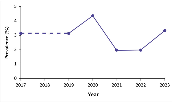
Objectives: This study aims to determine the prevalence and main imaging findings of PBC in women attending a tertiary breast imaging centre.
Method: A retrospective review of mammography and ultrasound imaging findings of female patients with histologically proven PBC, referred to a tertiary breast imaging centre over a 5-year period, was conducted.
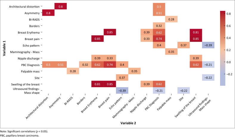
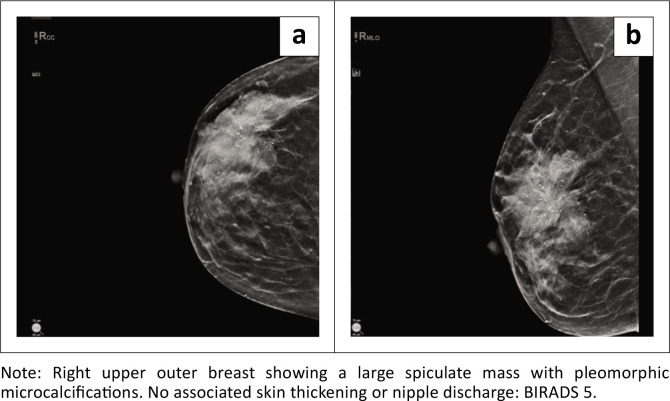
Results: The study included 102 female patients with a mean age of 53.8. Mammography detected masses in 93.02%, with calcifications in 41.2% and abnormal borders in 56.8%. Architectural distortion and asymmetry occurred in 27.5% and 28.4% respectively, both showing moderate correlation with PBC (r = 0.50, p = 0.009; r = 0.51, p = 0.0057). Ultrasound findings indicated irregular mass shapes (mean = 1.53), with hypoechoic patterns significantly associated with PBC (r = 0.40, p = 0.0013). Correlation analysis revealed strong associations between PBC and breast pain (r = 0.74, p < 0.0001), and erythema (r = 0.62, p < 0.0001). There was no significant association between the mammography and ultrasound findings (p = 0.495).
Conclusion: The findings underscore the value of using mammography and ultrasound in the diagnosis of PBC, as the two modalities offer complementary information.
Contribution: There is a paucity of data on the radiological findings of PBC in Africa. The current study prevalence mirrors global trends, highlighting the importance of ongoing surveillance and diagnostic accuracy.
期刊介绍:
The SA Journal of Radiology is the official journal of the Radiological Society of South Africa and the Professional Association of Radiologists in South Africa and Namibia. The SA Journal of Radiology is a general diagnostic radiological journal which carries original research and review articles, pictorial essays, case reports, letters, editorials, radiological practice and other radiological articles.

 求助内容:
求助内容: 应助结果提醒方式:


