Daniel Vogele, Janine Akbulut, Franziska Müller-Reichart, Aleš Janda, Henner Morbach, Hermann J Girschick, Matthias C Schaal, Meinrad Beer, Clemens Benoit
{"title":"The impact of sarcopenia on the progression of chronic non-bacterial osteomyelitis.","authors":"Daniel Vogele, Janine Akbulut, Franziska Müller-Reichart, Aleš Janda, Henner Morbach, Hermann J Girschick, Matthias C Schaal, Meinrad Beer, Clemens Benoit","doi":"10.1186/s13244-025-02001-w","DOIUrl":null,"url":null,"abstract":"<p><strong>Objectives: </strong>Chronic non-bacterial osteomyelitis (CNO) is the most common autoinflammatory bone disease in children and adolescents. This study investigated the progression of CNO lesions during therapy and the potential impact of sarcopenia on disease progression, utilizing routine MRI.</p><p><strong>Methods: </strong>A retrospective analysis of MRI examinations was conducted on 29 children and adolescents with CNO. CNO lesions were segmented. Sarcopenia was assessed using the total psoas muscle index (PMI) at lumbar vertebral levels L3/4 and L4/5. Measurements were taken at four time points during the disease course (T1: baseline, T2-T4: follow-up). Based on the PMI, patients were classified as sarcopenic or non-sarcopenic, and the progression of CNO lesions and the impact of sarcopenia were analyzed.</p><p><strong>Results: </strong>A total of 29 patients, aged 1-16 years, were included in the study, with 13 males and 16 females. Patients with sarcopenia had a significantly larger mean lesion area (868.95 mm<sup>2</sup>, SD = 684.49) compared to those without sarcopenia (636.11 mm<sup>2</sup>, SD = 832.41); p = 0.042, d = 0.4). The comparison between the two patient groups revealed a consistently lower percentage reduction in lesion size for the sarcopenic patients at all time points. Notably, the difference between T1 and T3 was statistically significant (p = 0.045, d = 0.82).</p><p><strong>Conclusion: </strong>The present study indicates that sarcopenia may serve as a negative prognostic factor in the treatment of CNO. Incorporating sarcopenia assessment as an additional parameter in routine whole-body MRI examinations could enhance the evaluation process.</p><p><strong>Critical relevance statement: </strong>Sarcopenia can be assessed using routine whole-body MRI in patients with CNO and may serve as a negative prognostic factor, potentially enhancing the evaluation process.</p><p><strong>Key points: </strong>Whole-body MRI is crucial for diagnosing and monitoring CNO. Routine whole-body MRI in CNO patients can also be used to assess sarcopenia as an additional parameter. Sarcopenia may act as a negative prognostic factor in CNO treatment, potentially improving the evaluation process.</p>","PeriodicalId":13639,"journal":{"name":"Insights into Imaging","volume":"16 1","pages":"117"},"PeriodicalIF":4.5000,"publicationDate":"2025-06-04","publicationTypes":"Journal Article","fieldsOfStudy":null,"isOpenAccess":false,"openAccessPdf":"https://www.ncbi.nlm.nih.gov/pmc/articles/PMC12137865/pdf/","citationCount":"0","resultStr":null,"platform":"Semanticscholar","paperid":null,"PeriodicalName":"Insights into Imaging","FirstCategoryId":"3","ListUrlMain":"https://doi.org/10.1186/s13244-025-02001-w","RegionNum":2,"RegionCategory":"医学","ArticlePicture":[],"TitleCN":null,"AbstractTextCN":null,"PMCID":null,"EPubDate":"","PubModel":"","JCR":"Q1","JCRName":"RADIOLOGY, NUCLEAR MEDICINE & MEDICAL IMAGING","Score":null,"Total":0}
引用次数: 0
Abstract
Objectives: Chronic non-bacterial osteomyelitis (CNO) is the most common autoinflammatory bone disease in children and adolescents. This study investigated the progression of CNO lesions during therapy and the potential impact of sarcopenia on disease progression, utilizing routine MRI.
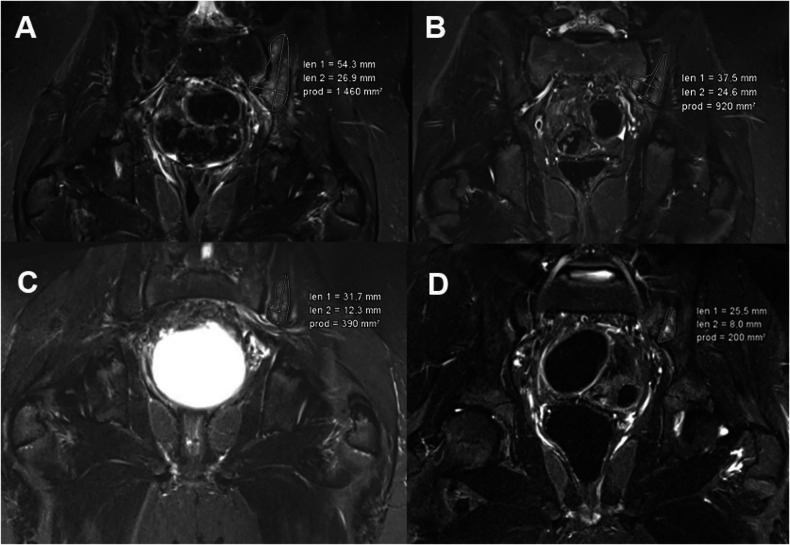
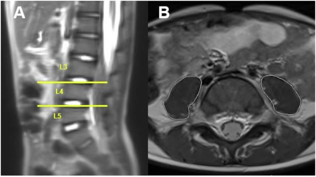
Methods: A retrospective analysis of MRI examinations was conducted on 29 children and adolescents with CNO. CNO lesions were segmented. Sarcopenia was assessed using the total psoas muscle index (PMI) at lumbar vertebral levels L3/4 and L4/5. Measurements were taken at four time points during the disease course (T1: baseline, T2-T4: follow-up). Based on the PMI, patients were classified as sarcopenic or non-sarcopenic, and the progression of CNO lesions and the impact of sarcopenia were analyzed.
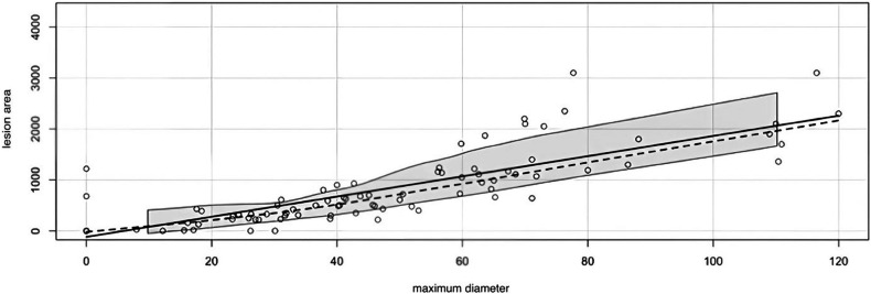
Results: A total of 29 patients, aged 1-16 years, were included in the study, with 13 males and 16 females. Patients with sarcopenia had a significantly larger mean lesion area (868.95 mm2, SD = 684.49) compared to those without sarcopenia (636.11 mm2, SD = 832.41); p = 0.042, d = 0.4). The comparison between the two patient groups revealed a consistently lower percentage reduction in lesion size for the sarcopenic patients at all time points. Notably, the difference between T1 and T3 was statistically significant (p = 0.045, d = 0.82).
Conclusion: The present study indicates that sarcopenia may serve as a negative prognostic factor in the treatment of CNO. Incorporating sarcopenia assessment as an additional parameter in routine whole-body MRI examinations could enhance the evaluation process.
Critical relevance statement: Sarcopenia can be assessed using routine whole-body MRI in patients with CNO and may serve as a negative prognostic factor, potentially enhancing the evaluation process.
Key points: Whole-body MRI is crucial for diagnosing and monitoring CNO. Routine whole-body MRI in CNO patients can also be used to assess sarcopenia as an additional parameter. Sarcopenia may act as a negative prognostic factor in CNO treatment, potentially improving the evaluation process.
期刊介绍:
Insights into Imaging (I³) is a peer-reviewed open access journal published under the brand SpringerOpen. All content published in the journal is freely available online to anyone, anywhere!
I³ continuously updates scientific knowledge and progress in best-practice standards in radiology through the publication of original articles and state-of-the-art reviews and opinions, along with recommendations and statements from the leading radiological societies in Europe.
Founded by the European Society of Radiology (ESR), I³ creates a platform for educational material, guidelines and recommendations, and a forum for topics of controversy.
A balanced combination of review articles, original papers, short communications from European radiological congresses and information on society matters makes I³ an indispensable source for current information in this field.
I³ is owned by the ESR, however authors retain copyright to their article according to the Creative Commons Attribution License (see Copyright and License Agreement). All articles can be read, redistributed and reused for free, as long as the author of the original work is cited properly.
The open access fees (article-processing charges) for this journal are kindly sponsored by ESR for all Members.
The journal went open access in 2012, which means that all articles published since then are freely available online.

 求助内容:
求助内容: 应助结果提醒方式:


