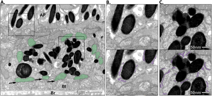
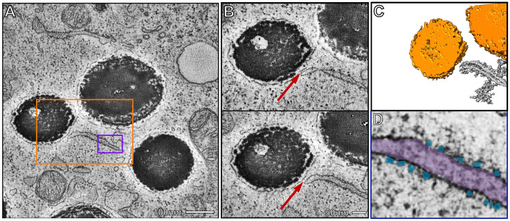
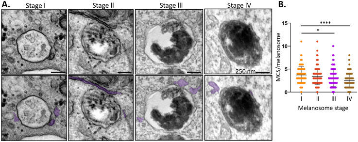
{"title":"Identification of ER:Melanosome Membrane Contact Sites in the Retinal Pigment Epithelium.","authors":"T Burgoyne, D Doncheva, E R Eden","doi":"10.1177/25152564251340949","DOIUrl":null,"url":null,"abstract":"<p><p>The retinal pigment epithelium (RPE) forms a monolayer of cells at the blood:retina interface that plays important roles for photoreceptor renewal and function and is central to retinal health. RPE pigment is provided by melanin-containing melanosomes which offer protection against light and oxidative stress. Melanosome migration into the apical processes of the RPE following light onset is thought to contribute to preventing retinal degeneration with age, though the mechanism is not yet clear. Melanosomes are transported along microtubules to the apical surface where they are transferred to actin filaments within the apical processes. Melanosomes are lysosome-related organelles derived from endosomes and endosome transport along microtubules is heavily influenced by the endoplasmic reticulum (ER) through ER:endosome contact sites. Here we describe extensive connection between the ER and melanosomes in the RPE. We further show, in skin melanocytes, that the ER forms contact sites with all stages of melanosome maturation, but ER contact is reduced as melanosomes mature. Finally, we identify tripartite contact sites between the ER, melanosomes and mitochondria in both RPE tissue and cellular models, suggesting that the ER may influence melanosome biogenesis, maturation and interaction with mitochondria.</p>","PeriodicalId":101304,"journal":{"name":"Contact (Thousand Oaks (Ventura County, Calif.))","volume":"8 ","pages":"25152564251340949"},"PeriodicalIF":0.0000,"publicationDate":"2025-06-02","publicationTypes":"Journal Article","fieldsOfStudy":null,"isOpenAccess":false,"openAccessPdf":"https://www.ncbi.nlm.nih.gov/pmc/articles/PMC12130655/pdf/","citationCount":"0","resultStr":null,"platform":"Semanticscholar","paperid":null,"PeriodicalName":"Contact (Thousand Oaks (Ventura County, Calif.))","FirstCategoryId":"1085","ListUrlMain":"https://doi.org/10.1177/25152564251340949","RegionNum":0,"RegionCategory":null,"ArticlePicture":[],"TitleCN":null,"AbstractTextCN":null,"PMCID":null,"EPubDate":"2025/1/1 0:00:00","PubModel":"eCollection","JCR":"","JCRName":"","Score":null,"Total":0}
引用次数: 0
Abstract
The retinal pigment epithelium (RPE) forms a monolayer of cells at the blood:retina interface that plays important roles for photoreceptor renewal and function and is central to retinal health. RPE pigment is provided by melanin-containing melanosomes which offer protection against light and oxidative stress. Melanosome migration into the apical processes of the RPE following light onset is thought to contribute to preventing retinal degeneration with age, though the mechanism is not yet clear. Melanosomes are transported along microtubules to the apical surface where they are transferred to actin filaments within the apical processes. Melanosomes are lysosome-related organelles derived from endosomes and endosome transport along microtubules is heavily influenced by the endoplasmic reticulum (ER) through ER:endosome contact sites. Here we describe extensive connection between the ER and melanosomes in the RPE. We further show, in skin melanocytes, that the ER forms contact sites with all stages of melanosome maturation, but ER contact is reduced as melanosomes mature. Finally, we identify tripartite contact sites between the ER, melanosomes and mitochondria in both RPE tissue and cellular models, suggesting that the ER may influence melanosome biogenesis, maturation and interaction with mitochondria.

 求助内容:
求助内容: 应助结果提醒方式:


