{"title":"Intratumoral and Peritumoral Radiomics Based on DCE-MRI for Prediction of Microvascular Invasion Grading in Solitary Hepatocellular Carcinoma (≤3 cm).","authors":"Yinqiao Li, Helin Li, Yayuan Feng, Lun Lu, Juan Zhang, Ningyang Jia","doi":"10.2147/JHC.S519578","DOIUrl":null,"url":null,"abstract":"<p><strong>Purpose: </strong>To explore the application value of clinical indicators, radiological features, and magnetic resonance imaging (MRI) radiomics to predict the grading of MVI in nodular hepatocellular carcinoma (≤3cm).</p><p><strong>Methods: </strong>A total of 131 patients with hepatocellular carcinoma (HCC) and confirmed microvascular invasion (MVI) who underwent surgical resection between January 2016 and December 2022 were retrospectively analyzed. A clinical-radiological (CR) model was constructed using independent risk factors identified by logistic regression. Radiomics models based on MRI (arterial phase, portal venous phase, delayed phase) across various regions (AVDP<sub>intra</sub>, AVDP<sub>intra+peri3mm</sub>, AVDP<sub>intra+peri5mm</sub>, AVDP<sub>intra+peri10mm</sub>) were developed using the Logistic Regression (LR) classifiers. The optimal radiomics model was subsequently integrated with the CR model to construct a combined clinical-radiological-radiomics (CRR) model. Model performance was assessed using the area under the curve (AUC).</p><p><strong>Results: </strong>Non-smooth margin and intratumoral artery were risk factors for MVI grading. The combined CRR model demonstrated the best predictive performance, with AUCs of 0.907 and 0.917 in the training and testing sets, respectively. Compared with the CR model alone, the CRR model showed a statistically significant improvement (p = 0.008, DeLong test).</p><p><strong>Conclusion: </strong>The AVDP<sub>intra+peri3mm</sub> model based on MRI radiomics demonstrates good predictive performance in predicting MVI grading in HCC (≤3cm). Combining features from the CR model with those of the AVDP<sub>intra+peri3mm</sub> model to construct the CRR model further enhances the prediction of MVI grading.</p>","PeriodicalId":15906,"journal":{"name":"Journal of Hepatocellular Carcinoma","volume":"12 ","pages":"1083-1095"},"PeriodicalIF":3.4000,"publicationDate":"2025-05-30","publicationTypes":"Journal Article","fieldsOfStudy":null,"isOpenAccess":false,"openAccessPdf":"https://www.ncbi.nlm.nih.gov/pmc/articles/PMC12132667/pdf/","citationCount":"0","resultStr":null,"platform":"Semanticscholar","paperid":null,"PeriodicalName":"Journal of Hepatocellular Carcinoma","FirstCategoryId":"3","ListUrlMain":"https://doi.org/10.2147/JHC.S519578","RegionNum":3,"RegionCategory":"医学","ArticlePicture":[],"TitleCN":null,"AbstractTextCN":null,"PMCID":null,"EPubDate":"2025/1/1 0:00:00","PubModel":"eCollection","JCR":"Q2","JCRName":"ONCOLOGY","Score":null,"Total":0}
引用次数: 0
Abstract
Purpose: To explore the application value of clinical indicators, radiological features, and magnetic resonance imaging (MRI) radiomics to predict the grading of MVI in nodular hepatocellular carcinoma (≤3cm).
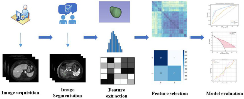
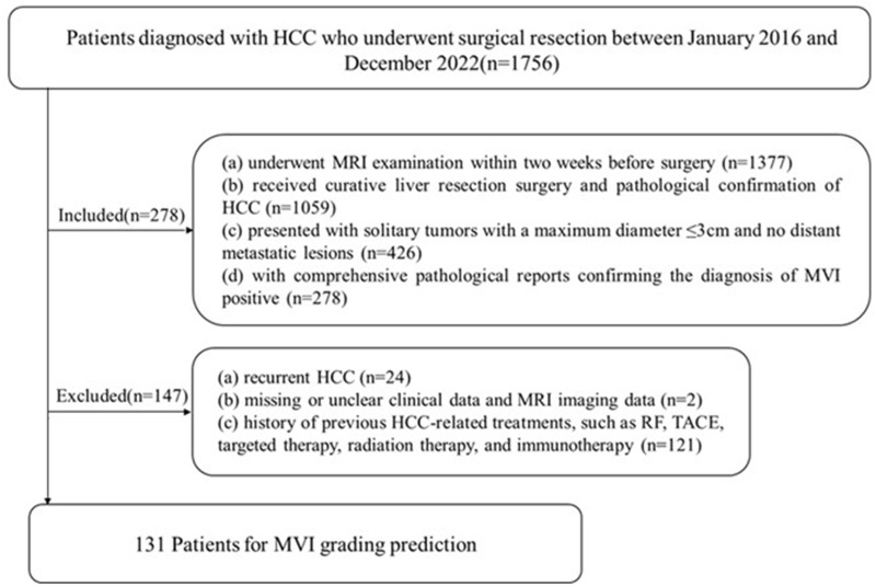
Methods: A total of 131 patients with hepatocellular carcinoma (HCC) and confirmed microvascular invasion (MVI) who underwent surgical resection between January 2016 and December 2022 were retrospectively analyzed. A clinical-radiological (CR) model was constructed using independent risk factors identified by logistic regression. Radiomics models based on MRI (arterial phase, portal venous phase, delayed phase) across various regions (AVDPintra, AVDPintra+peri3mm, AVDPintra+peri5mm, AVDPintra+peri10mm) were developed using the Logistic Regression (LR) classifiers. The optimal radiomics model was subsequently integrated with the CR model to construct a combined clinical-radiological-radiomics (CRR) model. Model performance was assessed using the area under the curve (AUC).
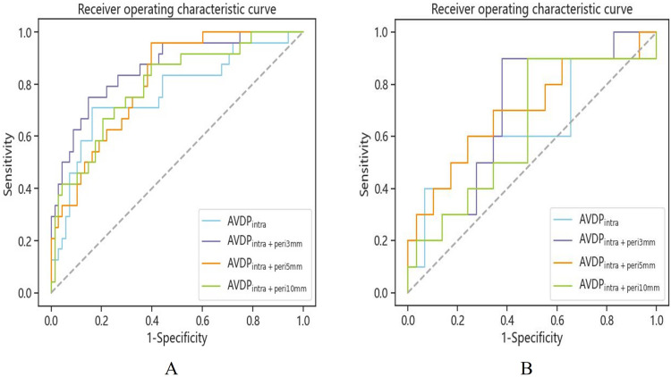
Results: Non-smooth margin and intratumoral artery were risk factors for MVI grading. The combined CRR model demonstrated the best predictive performance, with AUCs of 0.907 and 0.917 in the training and testing sets, respectively. Compared with the CR model alone, the CRR model showed a statistically significant improvement (p = 0.008, DeLong test).
Conclusion: The AVDPintra+peri3mm model based on MRI radiomics demonstrates good predictive performance in predicting MVI grading in HCC (≤3cm). Combining features from the CR model with those of the AVDPintra+peri3mm model to construct the CRR model further enhances the prediction of MVI grading.
 求助内容:
求助内容: 应助结果提醒方式:


