Ronald Seidel, Konrad Handrich, Marie Albéric, Jonathan Perrin, Derk Joester, Yael Politi and Luca Bertinetti
{"title":"Comparative structural analysis of stereom polymorphs in the sea urchin test†","authors":"Ronald Seidel, Konrad Handrich, Marie Albéric, Jonathan Perrin, Derk Joester, Yael Politi and Luca Bertinetti","doi":"10.1039/D5FD00033E","DOIUrl":null,"url":null,"abstract":"<p >The fenestrated ultrastructure of the sea urchin endoskeleton has attracted the attention of researchers in different fields due to its morphological complexity and crystallographic properties. Microscopic calcitic trabeculae form an intricate bicontinuous network, called the stereom. The stereom exhibits a wide variation of pore patterns, but is essentially a single calcite crystal (mono-crystalline). The polymorphism and crystal orientation in the skeletons of sea urchins have both been previously extensively described, mostly for taxonomical reasons and for mechanical studies. Moreover, while the resemblance of the stereom architecture to constant-mean-curvature (CMC) structures has been pointed out, a quantitative description and critical analysis is still lacking. Here, we use synchrotron micro-computed tomography to capture the three-dimensional (3D) architecture of the skeletal stereom in sea urchins for morphological quantification. By characterising the different stereom types, we define a data processing pipeline that allows inter-individual and interspecies comparison of stereom architectures, with implications for sea urchin taxonomy, mechanics, and skeletal growth. We further show that the various stereom morphologies are bicontinuous CMC surfaces that are unconstrained by crystallography. Our results highlight the properties of the soft tissue filling the stereom pore space in defining the shape of sea urchin biocalcite.</p>","PeriodicalId":49075,"journal":{"name":"Faraday Discussions","volume":"261 ","pages":" 340-358"},"PeriodicalIF":3.1000,"publicationDate":"2025-04-15","publicationTypes":"Journal Article","fieldsOfStudy":null,"isOpenAccess":false,"openAccessPdf":"https://pubs.rsc.org/en/content/articlepdf/2025/fd/d5fd00033e?page=search","citationCount":"0","resultStr":null,"platform":"Semanticscholar","paperid":null,"PeriodicalName":"Faraday Discussions","FirstCategoryId":"92","ListUrlMain":"https://pubs.rsc.org/en/content/articlelanding/2025/fd/d5fd00033e","RegionNum":3,"RegionCategory":"化学","ArticlePicture":[],"TitleCN":null,"AbstractTextCN":null,"PMCID":null,"EPubDate":"","PubModel":"","JCR":"Q2","JCRName":"Chemistry","Score":null,"Total":0}
引用次数: 0
Abstract
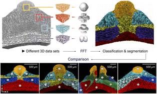
The fenestrated ultrastructure of the sea urchin endoskeleton has attracted the attention of researchers in different fields due to its morphological complexity and crystallographic properties. Microscopic calcitic trabeculae form an intricate bicontinuous network, called the stereom. The stereom exhibits a wide variation of pore patterns, but is essentially a single calcite crystal (mono-crystalline). The polymorphism and crystal orientation in the skeletons of sea urchins have both been previously extensively described, mostly for taxonomical reasons and for mechanical studies. Moreover, while the resemblance of the stereom architecture to constant-mean-curvature (CMC) structures has been pointed out, a quantitative description and critical analysis is still lacking. Here, we use synchrotron micro-computed tomography to capture the three-dimensional (3D) architecture of the skeletal stereom in sea urchins for morphological quantification. By characterising the different stereom types, we define a data processing pipeline that allows inter-individual and interspecies comparison of stereom architectures, with implications for sea urchin taxonomy, mechanics, and skeletal growth. We further show that the various stereom morphologies are bicontinuous CMC surfaces that are unconstrained by crystallography. Our results highlight the properties of the soft tissue filling the stereom pore space in defining the shape of sea urchin biocalcite.
 求助内容:
求助内容: 应助结果提醒方式:


