Qizheng Wang, Xiaoying Xing, Zixian Zhang, Xiaoxi Ji, Shipei He, Yuxin Yang, Jiajia Xu, Qiang Zhao, Ning Lang
{"title":"Added value of 3D fast-field-echo (FRACTURE) sequences for cervical spondylosis diagnosis: a prospective multi-reader non-inferiority study.","authors":"Qizheng Wang, Xiaoying Xing, Zixian Zhang, Xiaoxi Ji, Shipei He, Yuxin Yang, Jiajia Xu, Qiang Zhao, Ning Lang","doi":"10.1186/s13244-025-01997-5","DOIUrl":null,"url":null,"abstract":"<p><strong>Objectives: </strong>To assess the potential of fast field echo resembling a CT using restricted echo-spacing (FRACTURE) sequence to enhance conventional MRI in detecting bone abnormalities of cervical spondylosis.</p><p><strong>Materials and methods: </strong>137 consecutive patients with cervical spondylosis who underwent clinically indicated paired CT and MRI within 2 weeks between January and June 2024. After routine MRI, the 3D-FRACTURE sequences were performed. Three radiologists independently evaluated the data during three sessions: (1) CT with consensus, (2) routine MRI, and (3) FRACTURE, with a 4-week interval between sessions. Assessments included osteophytes, bony foraminal stenosis, posterior longitudinal ligament ossification (OPLL), their anatomical location, and diagnostic confidence, using CT as the reference standard. Inter- and intra-reader reproducibility was assessed using multi-rater Fleiss κ and the intraclass correlation coefficient (ICC), respectively. The non-inferiority assessment compared routine MRI/FRACTURE and CT diagnoses using a relative reduction margin of 0.5.</p><p><strong>Results: </strong>The study sample comprised 82 males and 55 females (age 56.9 ± 9.8 years). ICC indicated good to excellent inter-rater reliability for FRACTURE (osteophytes: ICC, 0.83-1.00; OPLL: ICC, 0.73-0.92; bony foraminal stenosis: ICC, 0.76-0.98), which was superior to conventional MRI (most ICC values < 0.7). The diagnostic confidence by FRACTURE sequences was significantly higher than by routine MRI (p < 0.001). Non-inferiority analysis demonstrated that FRACTURE and CT detection were similar for osteophyte, bony foraminal stenosis, and OPLL within a margin of 0.5.</p><p><strong>Conclusion: </strong>The FRACTURE sequence demonstrated comparable performance to CT in bone abnormalities detection in cervical spondylosis, superior to the routine MRI protocol.</p><p><strong>Critical relevance statement: </strong>The FRACTURE sequence addresses the limitations of conventional MRI in evaluating bone abnormalities, potentially minimizing radiation exposure and streamlining the diagnostic process for patients.</p><p><strong>Key points: </strong>MRI has advantages in the evaluation of cervical spondylosis, but is still insufficient in bone abnormalities evaluation. The FRACTURE sequence performed comparably to CT in bone abnormalities detection in cervical spondylosis. MRI with FRACTURE sequences may provide a non-ionizing method for assessing cervical spondylosis in some clinical settings.</p>","PeriodicalId":13639,"journal":{"name":"Insights into Imaging","volume":"16 1","pages":"114"},"PeriodicalIF":4.5000,"publicationDate":"2025-06-03","publicationTypes":"Journal Article","fieldsOfStudy":null,"isOpenAccess":false,"openAccessPdf":"https://www.ncbi.nlm.nih.gov/pmc/articles/PMC12133650/pdf/","citationCount":"0","resultStr":null,"platform":"Semanticscholar","paperid":null,"PeriodicalName":"Insights into Imaging","FirstCategoryId":"3","ListUrlMain":"https://doi.org/10.1186/s13244-025-01997-5","RegionNum":2,"RegionCategory":"医学","ArticlePicture":[],"TitleCN":null,"AbstractTextCN":null,"PMCID":null,"EPubDate":"","PubModel":"","JCR":"Q1","JCRName":"RADIOLOGY, NUCLEAR MEDICINE & MEDICAL IMAGING","Score":null,"Total":0}
引用次数: 0
Abstract
Objectives: To assess the potential of fast field echo resembling a CT using restricted echo-spacing (FRACTURE) sequence to enhance conventional MRI in detecting bone abnormalities of cervical spondylosis.
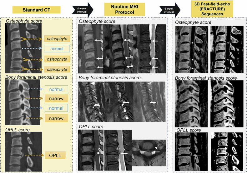
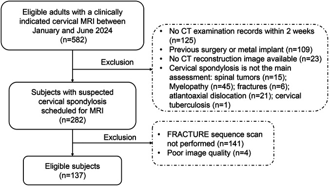
Materials and methods: 137 consecutive patients with cervical spondylosis who underwent clinically indicated paired CT and MRI within 2 weeks between January and June 2024. After routine MRI, the 3D-FRACTURE sequences were performed. Three radiologists independently evaluated the data during three sessions: (1) CT with consensus, (2) routine MRI, and (3) FRACTURE, with a 4-week interval between sessions. Assessments included osteophytes, bony foraminal stenosis, posterior longitudinal ligament ossification (OPLL), their anatomical location, and diagnostic confidence, using CT as the reference standard. Inter- and intra-reader reproducibility was assessed using multi-rater Fleiss κ and the intraclass correlation coefficient (ICC), respectively. The non-inferiority assessment compared routine MRI/FRACTURE and CT diagnoses using a relative reduction margin of 0.5.
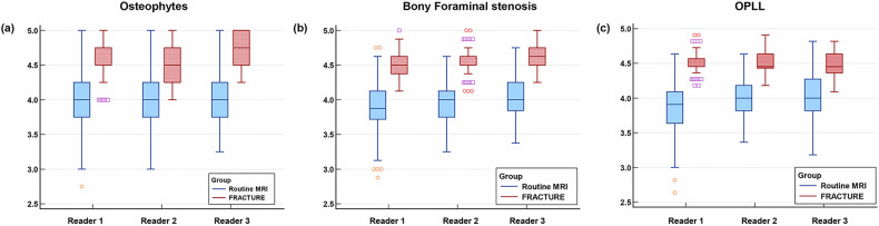
Results: The study sample comprised 82 males and 55 females (age 56.9 ± 9.8 years). ICC indicated good to excellent inter-rater reliability for FRACTURE (osteophytes: ICC, 0.83-1.00; OPLL: ICC, 0.73-0.92; bony foraminal stenosis: ICC, 0.76-0.98), which was superior to conventional MRI (most ICC values < 0.7). The diagnostic confidence by FRACTURE sequences was significantly higher than by routine MRI (p < 0.001). Non-inferiority analysis demonstrated that FRACTURE and CT detection were similar for osteophyte, bony foraminal stenosis, and OPLL within a margin of 0.5.
Conclusion: The FRACTURE sequence demonstrated comparable performance to CT in bone abnormalities detection in cervical spondylosis, superior to the routine MRI protocol.
Critical relevance statement: The FRACTURE sequence addresses the limitations of conventional MRI in evaluating bone abnormalities, potentially minimizing radiation exposure and streamlining the diagnostic process for patients.
Key points: MRI has advantages in the evaluation of cervical spondylosis, but is still insufficient in bone abnormalities evaluation. The FRACTURE sequence performed comparably to CT in bone abnormalities detection in cervical spondylosis. MRI with FRACTURE sequences may provide a non-ionizing method for assessing cervical spondylosis in some clinical settings.
期刊介绍:
Insights into Imaging (I³) is a peer-reviewed open access journal published under the brand SpringerOpen. All content published in the journal is freely available online to anyone, anywhere!
I³ continuously updates scientific knowledge and progress in best-practice standards in radiology through the publication of original articles and state-of-the-art reviews and opinions, along with recommendations and statements from the leading radiological societies in Europe.
Founded by the European Society of Radiology (ESR), I³ creates a platform for educational material, guidelines and recommendations, and a forum for topics of controversy.
A balanced combination of review articles, original papers, short communications from European radiological congresses and information on society matters makes I³ an indispensable source for current information in this field.
I³ is owned by the ESR, however authors retain copyright to their article according to the Creative Commons Attribution License (see Copyright and License Agreement). All articles can be read, redistributed and reused for free, as long as the author of the original work is cited properly.
The open access fees (article-processing charges) for this journal are kindly sponsored by ESR for all Members.
The journal went open access in 2012, which means that all articles published since then are freely available online.

 求助内容:
求助内容: 应助结果提醒方式:


