Forensic age estimation of the knee and clavicle using post-mortem CT
IF 1
Q4 RADIOLOGY, NUCLEAR MEDICINE & MEDICAL IMAGING
引用次数: 0
Abstract
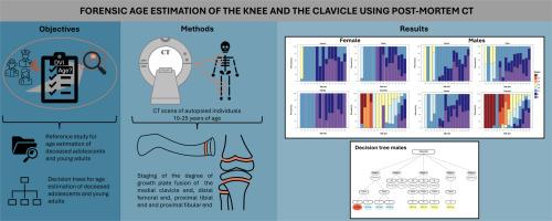
Age is crucial for constructing a biological profile to match data from missing persons in Disaster Victim Identification (DVI). For adolescents and young adults, methods often rely on bone end closures. Post-mortem computed tomography (PMCT) is a reliable tool for forensic age estimation in DVI cases. This study aimed to develop reference material for estimating the age of deceased adolescents and young adults using PMCT of the knee and clavicle in a Danish population. Results from the knee and clavicle were compared, and decision trees were created for final age estimates based on various bone combinations. The study included 221 individuals (57 females, 164 males) aged 10-25 years who underwent PMCT before medico-legal autopsy. Using the staging method by Schmeling et al. and Kellinghaus et al. ossification in the distal femoral, proximal tibial, proximal fibular, and medial clavicular epiphyses were evaluated. Large age intervals were observed for all bone stages, except for the earliest and latest clavicle stages. The clavicle stage distribution correlated well with chronological age, and reference values were consistent with previous studies. Individuals of younger age exhibited higher knee-bone stages than clavicle stages. Decision trees indicated that age intervals could be narrowed by combining clavicle stages with femur and fibula stages, though caution is required when intervals are too narrow. This study provides reference material for forensic age estimation in adolescents and young adults using PMCT of the knee and clavicle, relevant in DVI cases where limited body parts are available for analysis.
用死后CT对膝关节和锁骨的法医年龄估计
在灾害受害者身份识别(DVI)中,年龄对于构建与失踪人员数据相匹配的生物概况至关重要。对于青少年和年轻人,方法通常依赖于骨端闭合。死后计算机断层扫描(PMCT)是一个可靠的工具,法医年龄估计在DVI的情况下。本研究旨在为丹麦人群中使用膝关节和锁骨PMCT估计死亡青少年和年轻人的年龄提供参考资料。对膝关节和锁骨的结果进行比较,并根据不同的骨骼组合创建决策树以进行最终年龄估计。该研究包括221名10-25岁的个体(57名女性,164名男性),他们在法医尸检前接受了PMCT。采用Schmeling等人和Kellinghaus等人的分期方法,评估股骨远端、胫骨近端、腓骨近端和锁骨内侧骨骺的骨化情况。除了最早和最晚的锁骨期外,所有骨期的年龄间隔都很大。锁骨分期分布与实足年龄相关性较好,参考值与前人研究一致。年轻个体的膝骨阶段高于锁骨阶段。决策树显示年龄间隔可以通过结合锁骨分期与股骨和腓骨分期来缩小,但当间隔太窄时需要谨慎。本研究为使用膝关节和锁骨PMCT估计青少年和年轻人的法医年龄提供了参考资料,这与DVI病例中可用于分析的身体部位有限有关。
本文章由计算机程序翻译,如有差异,请以英文原文为准。
求助全文
约1分钟内获得全文
求助全文
来源期刊
Forensic Imaging
RADIOLOGY, NUCLEAR MEDICINE & MEDICAL IMAGING-
CiteScore
2.20
自引率
27.30%
发文量
39
 求助内容:
求助内容: 应助结果提醒方式:


