Elliot H Choi, Jose A Colmenarez, John D Hong, Kourosh Shahraki, Linxia Gu, Donny W Suh
{"title":"Finite element analysis of asymmetrical retinal hemorrhages in shaken baby syndrome.","authors":"Elliot H Choi, Jose A Colmenarez, John D Hong, Kourosh Shahraki, Linxia Gu, Donny W Suh","doi":"10.51329/mehdiophthal1514","DOIUrl":null,"url":null,"abstract":"<p><strong>Background: </strong>Despite the common association between bilateral retinal hemorrhage and shaken baby syndrome (SBS), unilateral retinal hemorrhage does not necessarily exclude this diagnosis. This study used computational simulations to elucidate the biomechanical phenomena within the eye under asymmetrical shaking forces.</p><p><strong>Methods: </strong>Finite element analysis (FEA) incorporating the vitreous, vitreoretinal interface, retinal layers, and retinal vessels was performed under asymmetrical shaking conditions. To assess the stress-strain response at the preretinal, intraretinal, and subretinal locations, we divided the retinal mesh into three equally spaced layers with an element height of 0.083 mm. The remaining space within the retina was filled with the vitreous humor and attached to it via the main retinal vessels extracted from a standard fundus image. The resulting changes in shear stress and intraocular pressure (IOP) were quantified.</p><p><strong>Results: </strong>The FEA model demonstrated that increasing the rotational radius from 10 cm to 14 cm or 17 cm led to a significant increase in shear stress and IOP across the vitreoretinal interface and within the retinal layers. Specifically, shear stress in the preretinal layer increased by 70.2% (8.0 kPa vs. 4.7 kPa), in the intraretinal layer by 20.0% (5.4 kPa vs. 4.5 kPa), and in the subretinal layer by 6.1% (3.5 kPa vs. 3.3 kPa). Simultaneously, IOP in the central region increased by 157.5% (39.4 mmHg vs. 15.3 mmHg) and in the posterior region by 162.3% (41.7 mmHg vs. 15.9 mmHg) when the rotational radius was increased to 17 cm from 10 cm. Increasing the rotational radius to 17 cm led to more pronounced changes in peak IOPs, with the central region showing a change of 39.4 mmHg and the posterior region a change of 41.7 mmHg. These results indicate a direct correlation between the rotational radius and the magnitude of IOP changes in the vitreous.</p><p><strong>Conclusions: </strong>These findings highlight the critical impact of rotational radius on the biomechanical forces exerted within the eye during asymmetrical shaking events, leading to variations in shear stress and IOP that could contribute to unilateral retinal hemorrhage in SBS. These insights reveal the complexity of diagnosing SBS and emphasize the need for careful consideration of the biomechanical evidence in patients presenting with asymmetrical or unilateral retinal hemorrhage.</p>","PeriodicalId":36524,"journal":{"name":"Medical Hypothesis, Discovery, and Innovation in Ophthalmology","volume":"14 1","pages":"231-238"},"PeriodicalIF":0.0000,"publicationDate":"2025-05-10","publicationTypes":"Journal Article","fieldsOfStudy":null,"isOpenAccess":false,"openAccessPdf":"https://www.ncbi.nlm.nih.gov/pmc/articles/PMC12121674/pdf/","citationCount":"0","resultStr":null,"platform":"Semanticscholar","paperid":null,"PeriodicalName":"Medical Hypothesis, Discovery, and Innovation in Ophthalmology","FirstCategoryId":"1085","ListUrlMain":"https://doi.org/10.51329/mehdiophthal1514","RegionNum":0,"RegionCategory":null,"ArticlePicture":[],"TitleCN":null,"AbstractTextCN":null,"PMCID":null,"EPubDate":"2025/1/1 0:00:00","PubModel":"eCollection","JCR":"Q2","JCRName":"Medicine","Score":null,"Total":0}
引用次数: 0
Abstract
Background: Despite the common association between bilateral retinal hemorrhage and shaken baby syndrome (SBS), unilateral retinal hemorrhage does not necessarily exclude this diagnosis. This study used computational simulations to elucidate the biomechanical phenomena within the eye under asymmetrical shaking forces.
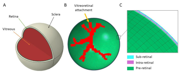
Methods: Finite element analysis (FEA) incorporating the vitreous, vitreoretinal interface, retinal layers, and retinal vessels was performed under asymmetrical shaking conditions. To assess the stress-strain response at the preretinal, intraretinal, and subretinal locations, we divided the retinal mesh into three equally spaced layers with an element height of 0.083 mm. The remaining space within the retina was filled with the vitreous humor and attached to it via the main retinal vessels extracted from a standard fundus image. The resulting changes in shear stress and intraocular pressure (IOP) were quantified.
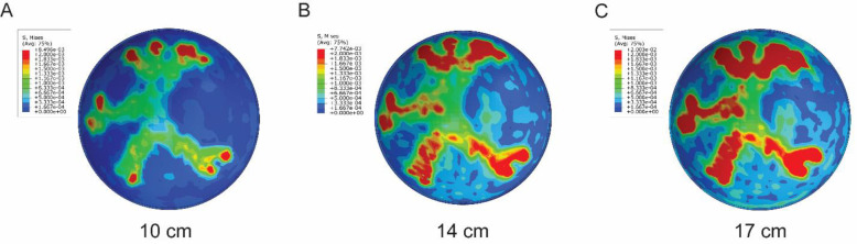
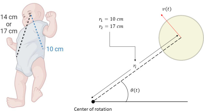
Results: The FEA model demonstrated that increasing the rotational radius from 10 cm to 14 cm or 17 cm led to a significant increase in shear stress and IOP across the vitreoretinal interface and within the retinal layers. Specifically, shear stress in the preretinal layer increased by 70.2% (8.0 kPa vs. 4.7 kPa), in the intraretinal layer by 20.0% (5.4 kPa vs. 4.5 kPa), and in the subretinal layer by 6.1% (3.5 kPa vs. 3.3 kPa). Simultaneously, IOP in the central region increased by 157.5% (39.4 mmHg vs. 15.3 mmHg) and in the posterior region by 162.3% (41.7 mmHg vs. 15.9 mmHg) when the rotational radius was increased to 17 cm from 10 cm. Increasing the rotational radius to 17 cm led to more pronounced changes in peak IOPs, with the central region showing a change of 39.4 mmHg and the posterior region a change of 41.7 mmHg. These results indicate a direct correlation between the rotational radius and the magnitude of IOP changes in the vitreous.
Conclusions: These findings highlight the critical impact of rotational radius on the biomechanical forces exerted within the eye during asymmetrical shaking events, leading to variations in shear stress and IOP that could contribute to unilateral retinal hemorrhage in SBS. These insights reveal the complexity of diagnosing SBS and emphasize the need for careful consideration of the biomechanical evidence in patients presenting with asymmetrical or unilateral retinal hemorrhage.

 求助内容:
求助内容: 应助结果提醒方式:


