Lingrui Cai, Craig Williamson, Andrew Nguyen, Emily Wittrup, Kayvan Najarian
{"title":"Adapting segment anything model for hematoma segmentation in traumatic brain injury.","authors":"Lingrui Cai, Craig Williamson, Andrew Nguyen, Emily Wittrup, Kayvan Najarian","doi":"10.1007/s44352-025-00011-4","DOIUrl":null,"url":null,"abstract":"<p><p>Hematoma segmentation in traumatic brain injury (TBI) is critical for accurate diagnosis and effective treatment planning. In this study, we evaluate various automated segmentation models, including stat-of-the-art architecture as benchmarks, and compare their performance with our proposed SAM-Adapter method for segmenting hematomas in brain CT scans. By incorporating the adapter into the vanilla SAM model, we address the challenges in medical imaging, which has very limited annotated datasets, enhancing model performance efficiency. We also find that domain-specific pre-processing, such as contrast adjustment, reduces the need for extensive pretraining, making the model more streamlined. And the model performance benefited with optimization and hyperparameter tuning. Our results demonstrate that the SAM-Adapter model achieved strong performance and reliability in identifying hematomas with Dice (72.34%), IoU (59.78%), 95% HD (5.57), sensitivity (75.39%) and specificity (99.73%). Inter-observer variability was assessed, revealing that the model's performance Dice (67.20%) was closely aligned with human expert agreement Dice (63.79%), suggesting its potential clinical utility. The external validation on the HemSeg-200 dataset, which contains 222 scans, demonstrates the robustness of our approach across diverse cases. These advancements in automatic segmentation hold promise for improving the accuracy and efficiency of TBI diagnosis, supporting clinical decision-making, and enhancing patient outcomes.</p><p><strong>Supplementary information: </strong>The online version contains supplementary material available at 10.1007/s44352-025-00011-4.</p>","PeriodicalId":520461,"journal":{"name":"Discover imaging","volume":"2 1","pages":"6"},"PeriodicalIF":0.0000,"publicationDate":"2025-01-01","publicationTypes":"Journal Article","fieldsOfStudy":null,"isOpenAccess":false,"openAccessPdf":"https://www.ncbi.nlm.nih.gov/pmc/articles/PMC12106135/pdf/","citationCount":"0","resultStr":null,"platform":"Semanticscholar","paperid":null,"PeriodicalName":"Discover imaging","FirstCategoryId":"1085","ListUrlMain":"https://doi.org/10.1007/s44352-025-00011-4","RegionNum":0,"RegionCategory":null,"ArticlePicture":[],"TitleCN":null,"AbstractTextCN":null,"PMCID":null,"EPubDate":"2025/5/26 0:00:00","PubModel":"Epub","JCR":"","JCRName":"","Score":null,"Total":0}
引用次数: 0
Abstract
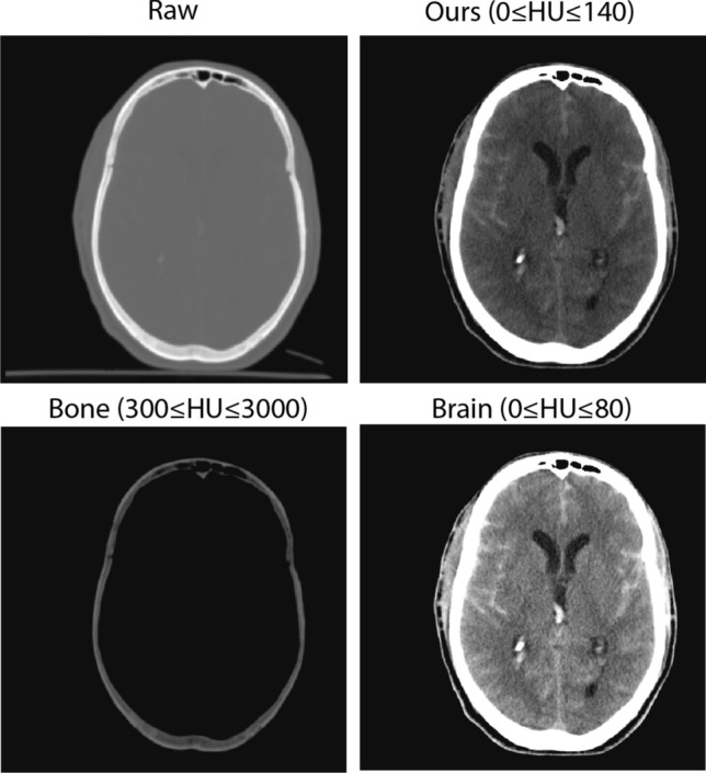
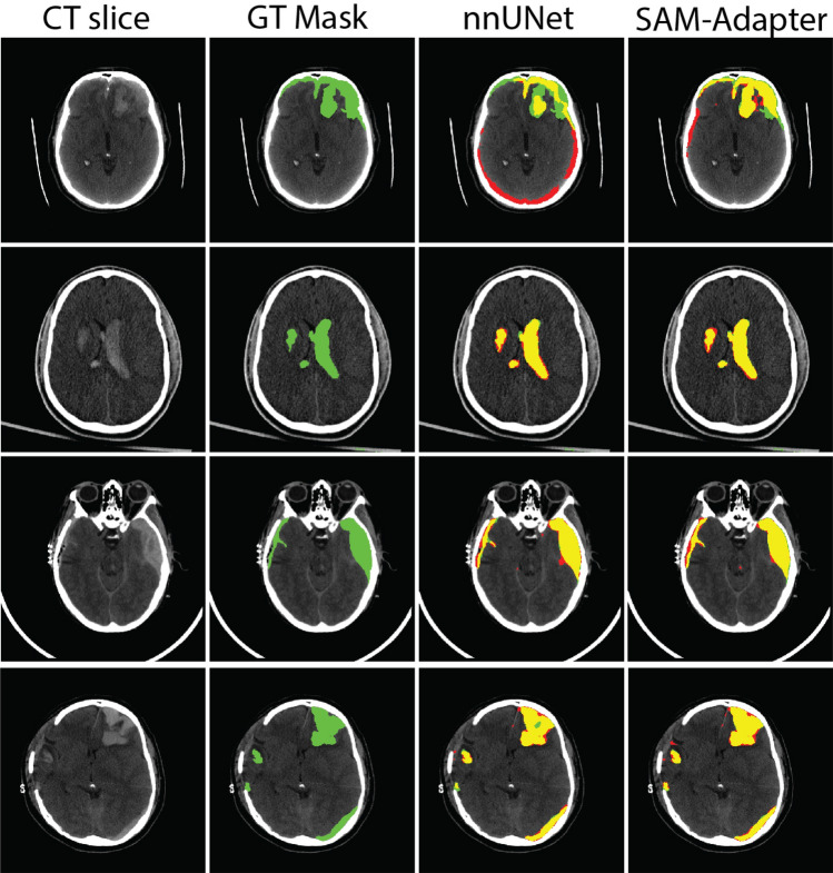
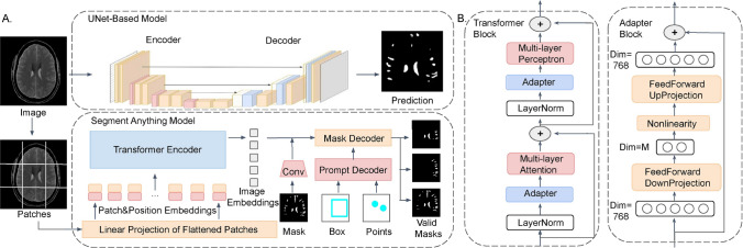
Hematoma segmentation in traumatic brain injury (TBI) is critical for accurate diagnosis and effective treatment planning. In this study, we evaluate various automated segmentation models, including stat-of-the-art architecture as benchmarks, and compare their performance with our proposed SAM-Adapter method for segmenting hematomas in brain CT scans. By incorporating the adapter into the vanilla SAM model, we address the challenges in medical imaging, which has very limited annotated datasets, enhancing model performance efficiency. We also find that domain-specific pre-processing, such as contrast adjustment, reduces the need for extensive pretraining, making the model more streamlined. And the model performance benefited with optimization and hyperparameter tuning. Our results demonstrate that the SAM-Adapter model achieved strong performance and reliability in identifying hematomas with Dice (72.34%), IoU (59.78%), 95% HD (5.57), sensitivity (75.39%) and specificity (99.73%). Inter-observer variability was assessed, revealing that the model's performance Dice (67.20%) was closely aligned with human expert agreement Dice (63.79%), suggesting its potential clinical utility. The external validation on the HemSeg-200 dataset, which contains 222 scans, demonstrates the robustness of our approach across diverse cases. These advancements in automatic segmentation hold promise for improving the accuracy and efficiency of TBI diagnosis, supporting clinical decision-making, and enhancing patient outcomes.
Supplementary information: The online version contains supplementary material available at 10.1007/s44352-025-00011-4.

 求助内容:
求助内容: 应助结果提醒方式:


