Andrea Zito, Francesco Burzotta, Cristina Aurigemma, Enrico Romagnoli, Lazzaro Paraggio, Francesco Fracassi, Mattia Lunardi, Luigi Cappannoli, Francesco Bianchini, Carlo Trani
{"title":"Intravascular imaging for percutaneous coronary intervention on bifurcation and unprotected left main lesions: a systematic review and meta-analysis.","authors":"Andrea Zito, Francesco Burzotta, Cristina Aurigemma, Enrico Romagnoli, Lazzaro Paraggio, Francesco Fracassi, Mattia Lunardi, Luigi Cappannoli, Francesco Bianchini, Carlo Trani","doi":"10.1136/openhrt-2024-003026","DOIUrl":null,"url":null,"abstract":"<p><strong>Background: </strong>The efficacy of intravascular imaging (IVI) guidance for percutaneous coronary intervention (PCI) represents a contemporary hot topic. PCI in patients with bifurcation coronary lesions and unprotected left main lesions offers specific challenges that, theoretically, may particularly benefit from IVI.</p><p><strong>Objective: </strong>To compare the clinical outcomes between IVI and angiography guidance for PCI in bifurcation and unprotected left main lesions.</p><p><strong>Methods: </strong>Randomised clinical trials (RCTs) comparing IVI (with either intravascular ultrasound or optical coherence tomography) with angiography to guide PCI in patients with bifurcation and unprotected left main lesions were searched in PubMed and Cochrane Central Register of Controlled Trials. Two investigators independently extracted study data. Risk ratios (RRs) were calculated using the random-effects model with inverse variance weighting and the 95% CIs with the modified Knapp-Hartung-Sidik-Jonkman method. The primary outcome was target vessel failure (TVF).</p><p><strong>Results: </strong>A total of seven RCTs were included, collecting data on 2494 patients in the analysis for bifurcation lesions and 1107 patients in the analysis for unprotected left main lesions. The mean follow-up duration ranged from 12 to 36 months. Compared with angiography guidance, IVI guidance significantly reduced TVF both in bifurcation lesions (RR 0.70, 95% CI 0.53 to 0.92) and unprotected left main lesions (RR 0.55, 95% CI 0.36 to 0.84). The number needed to treat to prevent one TVF with IVI was 27 in bifurcation lesions PCI and 11 in unprotected left main PCI.</p><p><strong>Conclusion: </strong>In patients undergoing PCI on bifurcation and unprotected left main lesions, IVI guidance significantly reduces the risk of TVF compared with angiography guidance.</p><p><strong>Prospero registration number: </strong>CRD42024580321.</p>","PeriodicalId":19505,"journal":{"name":"Open Heart","volume":"12 1","pages":""},"PeriodicalIF":2.8000,"publicationDate":"2025-05-27","publicationTypes":"Journal Article","fieldsOfStudy":null,"isOpenAccess":false,"openAccessPdf":"https://www.ncbi.nlm.nih.gov/pmc/articles/PMC12121598/pdf/","citationCount":"0","resultStr":null,"platform":"Semanticscholar","paperid":null,"PeriodicalName":"Open Heart","FirstCategoryId":"1085","ListUrlMain":"https://doi.org/10.1136/openhrt-2024-003026","RegionNum":0,"RegionCategory":null,"ArticlePicture":[],"TitleCN":null,"AbstractTextCN":null,"PMCID":null,"EPubDate":"","PubModel":"","JCR":"Q2","JCRName":"CARDIAC & CARDIOVASCULAR SYSTEMS","Score":null,"Total":0}
引用次数: 0
Abstract
Background: The efficacy of intravascular imaging (IVI) guidance for percutaneous coronary intervention (PCI) represents a contemporary hot topic. PCI in patients with bifurcation coronary lesions and unprotected left main lesions offers specific challenges that, theoretically, may particularly benefit from IVI.
Objective: To compare the clinical outcomes between IVI and angiography guidance for PCI in bifurcation and unprotected left main lesions.
Methods: Randomised clinical trials (RCTs) comparing IVI (with either intravascular ultrasound or optical coherence tomography) with angiography to guide PCI in patients with bifurcation and unprotected left main lesions were searched in PubMed and Cochrane Central Register of Controlled Trials. Two investigators independently extracted study data. Risk ratios (RRs) were calculated using the random-effects model with inverse variance weighting and the 95% CIs with the modified Knapp-Hartung-Sidik-Jonkman method. The primary outcome was target vessel failure (TVF).
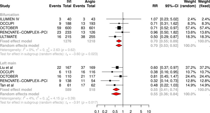
Results: A total of seven RCTs were included, collecting data on 2494 patients in the analysis for bifurcation lesions and 1107 patients in the analysis for unprotected left main lesions. The mean follow-up duration ranged from 12 to 36 months. Compared with angiography guidance, IVI guidance significantly reduced TVF both in bifurcation lesions (RR 0.70, 95% CI 0.53 to 0.92) and unprotected left main lesions (RR 0.55, 95% CI 0.36 to 0.84). The number needed to treat to prevent one TVF with IVI was 27 in bifurcation lesions PCI and 11 in unprotected left main PCI.
Conclusion: In patients undergoing PCI on bifurcation and unprotected left main lesions, IVI guidance significantly reduces the risk of TVF compared with angiography guidance.
期刊介绍:
Open Heart is an online-only, open access cardiology journal that aims to be “open” in many ways: open access (free access for all readers), open peer review (unblinded peer review) and open data (data sharing is encouraged). The goal is to ensure maximum transparency and maximum impact on research progress and patient care. The journal is dedicated to publishing high quality, peer reviewed medical research in all disciplines and therapeutic areas of cardiovascular medicine. Research is published across all study phases and designs, from study protocols to phase I trials to meta-analyses, including small or specialist studies. Opinionated discussions on controversial topics are welcomed. Open Heart aims to operate a fast submission and review process with continuous publication online, to ensure timely, up-to-date research is available worldwide. The journal adheres to a rigorous and transparent peer review process, and all articles go through a statistical assessment to ensure robustness of the analyses. Open Heart is an official journal of the British Cardiovascular Society.

 求助内容:
求助内容: 应助结果提醒方式:


