{"title":"A Performance Indicator for Optimizing Source–Detector Separation in Functional Near-Infrared Spectroscopy","authors":"Serhat Ilgaz Yoner, Gokhan Ertas","doi":"10.1002/ima.70113","DOIUrl":null,"url":null,"abstract":"<p>The performance of Functional Near-Infrared Spectroscopy (fNIRS) devices critically depends on the probe design, which affects signal quality, spatial and depth resolution, and data reliability. A critical component of probe separation is source-to-detector separation, which is defined as the distance between the light source and the detector. Optimizing this separation is essential for improving the signal-to-noise ratio (SNR) and sensitivity at depth (SAD). Larger separations enhance depth resolution, facilitating more accurate assessments of brain activity. Conversely, excessive separation may reduce SNR due to the lower light intensity received by the detector. In this study, a performance indicator was created to optimize separation by integrating the SNR and SAD. A probe was constructed that featured one light source and four detectors mounted on a mechanism that allowed for adjustable separations. A phantom mimicking brain tissue was used. Signals were recorded from the probe positioned on the phantom at various separations, employing light sources emitting light at wavelengths of 730, 800, and 850 nm, and optical power levels of 19, 26, 32, 38, and 44 mW. The SNR values for each separation were computed from the recorded signals, whereas the SAD values were obtained from existing literature. The performance indicator was developed as a weighted sum of SNR and SAD, normalized between 0 and 1, with higher values indicating enhanced probe performance due to optimized separation. The indicator is expected to improve the reliability of fNIRS data; however, further research involving diverse populations is required to validate its practical application.</p>","PeriodicalId":14027,"journal":{"name":"International Journal of Imaging Systems and Technology","volume":"35 4","pages":""},"PeriodicalIF":2.5000,"publicationDate":"2025-05-29","publicationTypes":"Journal Article","fieldsOfStudy":null,"isOpenAccess":false,"openAccessPdf":"https://onlinelibrary.wiley.com/doi/epdf/10.1002/ima.70113","citationCount":"0","resultStr":null,"platform":"Semanticscholar","paperid":null,"PeriodicalName":"International Journal of Imaging Systems and Technology","FirstCategoryId":"94","ListUrlMain":"https://onlinelibrary.wiley.com/doi/10.1002/ima.70113","RegionNum":4,"RegionCategory":"计算机科学","ArticlePicture":[],"TitleCN":null,"AbstractTextCN":null,"PMCID":null,"EPubDate":"","PubModel":"","JCR":"Q2","JCRName":"ENGINEERING, ELECTRICAL & ELECTRONIC","Score":null,"Total":0}
引用次数: 0
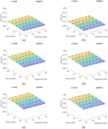
Abstract
The performance of Functional Near-Infrared Spectroscopy (fNIRS) devices critically depends on the probe design, which affects signal quality, spatial and depth resolution, and data reliability. A critical component of probe separation is source-to-detector separation, which is defined as the distance between the light source and the detector. Optimizing this separation is essential for improving the signal-to-noise ratio (SNR) and sensitivity at depth (SAD). Larger separations enhance depth resolution, facilitating more accurate assessments of brain activity. Conversely, excessive separation may reduce SNR due to the lower light intensity received by the detector. In this study, a performance indicator was created to optimize separation by integrating the SNR and SAD. A probe was constructed that featured one light source and four detectors mounted on a mechanism that allowed for adjustable separations. A phantom mimicking brain tissue was used. Signals were recorded from the probe positioned on the phantom at various separations, employing light sources emitting light at wavelengths of 730, 800, and 850 nm, and optical power levels of 19, 26, 32, 38, and 44 mW. The SNR values for each separation were computed from the recorded signals, whereas the SAD values were obtained from existing literature. The performance indicator was developed as a weighted sum of SNR and SAD, normalized between 0 and 1, with higher values indicating enhanced probe performance due to optimized separation. The indicator is expected to improve the reliability of fNIRS data; however, further research involving diverse populations is required to validate its practical application.
期刊介绍:
The International Journal of Imaging Systems and Technology (IMA) is a forum for the exchange of ideas and results relevant to imaging systems, including imaging physics and informatics. The journal covers all imaging modalities in humans and animals.
IMA accepts technically sound and scientifically rigorous research in the interdisciplinary field of imaging, including relevant algorithmic research and hardware and software development, and their applications relevant to medical research. The journal provides a platform to publish original research in structural and functional imaging.
The journal is also open to imaging studies of the human body and on animals that describe novel diagnostic imaging and analyses methods. Technical, theoretical, and clinical research in both normal and clinical populations is encouraged. Submissions describing methods, software, databases, replication studies as well as negative results are also considered.
The scope of the journal includes, but is not limited to, the following in the context of biomedical research:
Imaging and neuro-imaging modalities: structural MRI, functional MRI, PET, SPECT, CT, ultrasound, EEG, MEG, NIRS etc.;
Neuromodulation and brain stimulation techniques such as TMS and tDCS;
Software and hardware for imaging, especially related to human and animal health;
Image segmentation in normal and clinical populations;
Pattern analysis and classification using machine learning techniques;
Computational modeling and analysis;
Brain connectivity and connectomics;
Systems-level characterization of brain function;
Neural networks and neurorobotics;
Computer vision, based on human/animal physiology;
Brain-computer interface (BCI) technology;
Big data, databasing and data mining.

 求助内容:
求助内容: 应助结果提醒方式:


