Nathan Chaclas, Carter E Hall, Bernard D Horn, Richard S Davidson
{"title":"3D Mapping of Talocalcaneal Coalitions: An Aid to Surgical Approach and Excision.","authors":"Nathan Chaclas, Carter E Hall, Bernard D Horn, Richard S Davidson","doi":"10.1016/j.jposna.2025.100166","DOIUrl":null,"url":null,"abstract":"<p><strong>Background: </strong>The traditional approach to talocalcaneal tarsal coalition (TCC) excision is medial through the deltoid ligament. Unfortunately, there are few anatomic markers to guide the surgeon. Preoperative planning includes radiographs and advanced imaging; these currently provide little help guiding the excision. Our method of mapping the coalition on advanced imaging accurately defines where to make osteotomies in the operating room.</p><p><strong>Methods: </strong>A retrospective review was conducted of TCC patients with preoperative computed tomography (CT) at a single institution from 2010 to 2022. Three independent raters reported sagittal TCC length relative to the talus. Raters further quantified TCC coronal depth and height across distal, middle, and proximal thirds of the TCC relative to the sustentaculum talus. This guidance directs two osteotomy cuts through the coalition directly into the normal lateral subtalar joint for complete and accurate excision of the coalition. CT measurement inter-rater reliability was determined using intra-class correlation.</p><p><strong>Results: </strong>Twenty-seven patients (16 male), average age 13.9 ± 2.4, met study criteria. TCCs were located on the right lower extremity in 10/27 cases. Sixteen/twenty-seven coalitions were horizontal, 10/27 was down sloping, and 1/27 was upsloping relative to the joint line. Substantial agreement was achieved between three raters (mean average measures intraclass correlation 0.781). The mean coalition length in the sagittal plane was 21.2 ± 6.0 mm, covering 49.6 ± 23.2% of the talar length.</p><p><strong>Conclusion: </strong>This study describes a 3D preoperative mapping technique with high reproducibility among the present raters to resect the TCC with direct vision of the normal subtalar joints. Alternative approaches, such as obtaining CT imaging intraoperatively, expose the patient to increased radiation and anesthesia, incurring higher financial and time costs. We report a concise, readily applicable, and systematic method to map TCCs on preoperative CT and provide direct vision of the normal subtalar (talar and calcaneal) joints, as well as close to normal subtalar motion.</p><p><strong>Key concepts: </strong>(1)To date, very little in the way of intraoperative planning for TCCs has been proposed in the literature, even though advanced imaging has been widely used preoperatively.(2)Our method of mapping coalitions on preoperative CT may assist with intraoperative resection. Additionally, this method demonstrates the three-dimensional variety that can be expected in surgical excision of these coalitions.(3)Neither the medial to lateral depth nor the distance proximal from the sustentaculum talus was uniform as the coalitions were thickest centrally and tapered both proximally and distally.</p><p><strong>Level of evidence: </strong>IV.</p>","PeriodicalId":520850,"journal":{"name":"Journal of the Pediatric Orthopaedic Society of North America","volume":"11 ","pages":"100166"},"PeriodicalIF":0.0000,"publicationDate":"2025-03-07","publicationTypes":"Journal Article","fieldsOfStudy":null,"isOpenAccess":false,"openAccessPdf":"https://www.ncbi.nlm.nih.gov/pmc/articles/PMC12088112/pdf/","citationCount":"0","resultStr":null,"platform":"Semanticscholar","paperid":null,"PeriodicalName":"Journal of the Pediatric Orthopaedic Society of North America","FirstCategoryId":"1085","ListUrlMain":"https://doi.org/10.1016/j.jposna.2025.100166","RegionNum":0,"RegionCategory":null,"ArticlePicture":[],"TitleCN":null,"AbstractTextCN":null,"PMCID":null,"EPubDate":"2025/5/1 0:00:00","PubModel":"eCollection","JCR":"","JCRName":"","Score":null,"Total":0}
引用次数: 0
Abstract
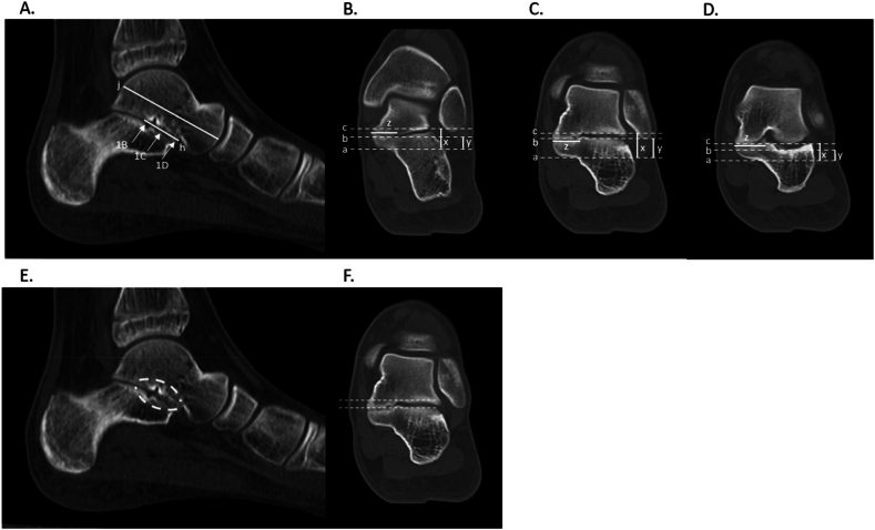
Background: The traditional approach to talocalcaneal tarsal coalition (TCC) excision is medial through the deltoid ligament. Unfortunately, there are few anatomic markers to guide the surgeon. Preoperative planning includes radiographs and advanced imaging; these currently provide little help guiding the excision. Our method of mapping the coalition on advanced imaging accurately defines where to make osteotomies in the operating room.
Methods: A retrospective review was conducted of TCC patients with preoperative computed tomography (CT) at a single institution from 2010 to 2022. Three independent raters reported sagittal TCC length relative to the talus. Raters further quantified TCC coronal depth and height across distal, middle, and proximal thirds of the TCC relative to the sustentaculum talus. This guidance directs two osteotomy cuts through the coalition directly into the normal lateral subtalar joint for complete and accurate excision of the coalition. CT measurement inter-rater reliability was determined using intra-class correlation.
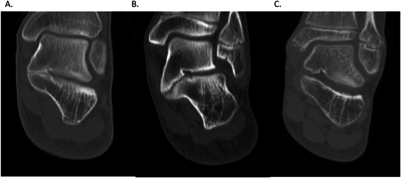
Results: Twenty-seven patients (16 male), average age 13.9 ± 2.4, met study criteria. TCCs were located on the right lower extremity in 10/27 cases. Sixteen/twenty-seven coalitions were horizontal, 10/27 was down sloping, and 1/27 was upsloping relative to the joint line. Substantial agreement was achieved between three raters (mean average measures intraclass correlation 0.781). The mean coalition length in the sagittal plane was 21.2 ± 6.0 mm, covering 49.6 ± 23.2% of the talar length.
Conclusion: This study describes a 3D preoperative mapping technique with high reproducibility among the present raters to resect the TCC with direct vision of the normal subtalar joints. Alternative approaches, such as obtaining CT imaging intraoperatively, expose the patient to increased radiation and anesthesia, incurring higher financial and time costs. We report a concise, readily applicable, and systematic method to map TCCs on preoperative CT and provide direct vision of the normal subtalar (talar and calcaneal) joints, as well as close to normal subtalar motion.
Key concepts: (1)To date, very little in the way of intraoperative planning for TCCs has been proposed in the literature, even though advanced imaging has been widely used preoperatively.(2)Our method of mapping coalitions on preoperative CT may assist with intraoperative resection. Additionally, this method demonstrates the three-dimensional variety that can be expected in surgical excision of these coalitions.(3)Neither the medial to lateral depth nor the distance proximal from the sustentaculum talus was uniform as the coalitions were thickest centrally and tapered both proximally and distally.

 求助内容:
求助内容: 应助结果提醒方式:


