Nabil K El Ayoubi, Ali Ismail, Georgio Sader, Nour Abi Chakra, Jad El Ahdab, Joseph Abboud, Samia J Khoury
{"title":"Retinal Optical Coherence Tomography Longitudinal Measures as Prognostic Biomarkers in Multiple Sclerosis: Systematic Review and Meta-Analysis.","authors":"Nabil K El Ayoubi, Ali Ismail, Georgio Sader, Nour Abi Chakra, Jad El Ahdab, Joseph Abboud, Samia J Khoury","doi":"10.1212/NXI.0000000000200416","DOIUrl":null,"url":null,"abstract":"<p><strong>Background and objectives: </strong>Optical coherence tomography (OCT) has emerged as a valuable marker for assessing inflammation and neuroaxonal degeneration in multiple sclerosis (MS). Although traditional markers such as brain atrophy and axonal loss are crucial for monitoring MS progression, their clinical application can be limited by various factors. This meta-analysis of longitudinal studies aims to assess the predictive value of OCT-derived retinal layer thickness thresholds for monitoring and predicting MS disease progression and cognitive decline.</p><p><strong>Methods: </strong>Our systematic review and meta-analysis followed Preferred Reporting Items for Systematic Reviews and Meta-analyses guidelines. A comprehensive systematic search was performed using electronic databases (PubMed, Embase, Web of Science, and Google Scholar) for longitudinal studies using Spectral Domain-OCT (SD-OCT) to assess retinal layer thickness and its predictive value for MS progression. Data were extracted on study design, OCT measurements, disability progression definitions, and clinical outcomes. We analyzed hazard ratios (HR) and odds ratios (OR) for associations between OCT-measured thresholds and disability progression, including physical and cognitive deterioration.</p><p><strong>Results: </strong>Our study included 14 longitudinal studies that met our inclusion criteria, 13 studies were included in our quantitative analysis, with a total of 3,683 participants. Baseline peripapillary retinal nerve fiber layer (pRNFL) thickness below 88 μm was significantly associated with increased risk of future disease progression and physical worsening measured by Expanded Disability Status Scale progression (HR = 2.376, <i>p</i> < 0.001; HR = 2.258, <i>p</i> < 0.001, respectively). The same was noted for ganglion cell-inner plexiform layer (GCIPL) thickness below 77 μm (HR = 2.751, <i>p</i> < 0.001 and HR = 2.66, <i>p</i> < 0.001, respectively). In addition, annualized rates of pRNFL thinning above 1.5 μm/y and GCIPL thinning above 1 μm/y also significantly predicted disease worsening (HR = 3.019, <i>p</i> = 0.005 and HR = 3.535, <i>p</i> < 0.001, respectively).</p><p><strong>Discussion: </strong>OCT-derived retinal layer thresholds, specifically a pRNFL thickness of ≤88 μm and a GCIPL thickness of ≤77 μm, are significantly associated with an increased risk of future MS disability progression. Furthermore, annual thinning rates of pRNFL >1.5 μm/y and GCIPL >1 μm/y demonstrate greater predictive power and are more clinically relevant for identifying individuals at high risk of both physical and cognitive disability progression outcomes. Further research is needed to standardize OCT thresholds and improve clinical use in treatment planning.</p>","PeriodicalId":19472,"journal":{"name":"Neurology® Neuroimmunology & Neuroinflammation","volume":"12 4","pages":"e200416"},"PeriodicalIF":7.5000,"publicationDate":"2025-07-01","publicationTypes":"Journal Article","fieldsOfStudy":null,"isOpenAccess":false,"openAccessPdf":"https://www.ncbi.nlm.nih.gov/pmc/articles/PMC12153945/pdf/","citationCount":"0","resultStr":null,"platform":"Semanticscholar","paperid":null,"PeriodicalName":"Neurology® Neuroimmunology & Neuroinflammation","FirstCategoryId":"3","ListUrlMain":"https://doi.org/10.1212/NXI.0000000000200416","RegionNum":1,"RegionCategory":"医学","ArticlePicture":[],"TitleCN":null,"AbstractTextCN":null,"PMCID":null,"EPubDate":"2025/5/27 0:00:00","PubModel":"Epub","JCR":"Q1","JCRName":"CLINICAL NEUROLOGY","Score":null,"Total":0}
引用次数: 0
Abstract
Background and objectives: Optical coherence tomography (OCT) has emerged as a valuable marker for assessing inflammation and neuroaxonal degeneration in multiple sclerosis (MS). Although traditional markers such as brain atrophy and axonal loss are crucial for monitoring MS progression, their clinical application can be limited by various factors. This meta-analysis of longitudinal studies aims to assess the predictive value of OCT-derived retinal layer thickness thresholds for monitoring and predicting MS disease progression and cognitive decline.
Methods: Our systematic review and meta-analysis followed Preferred Reporting Items for Systematic Reviews and Meta-analyses guidelines. A comprehensive systematic search was performed using electronic databases (PubMed, Embase, Web of Science, and Google Scholar) for longitudinal studies using Spectral Domain-OCT (SD-OCT) to assess retinal layer thickness and its predictive value for MS progression. Data were extracted on study design, OCT measurements, disability progression definitions, and clinical outcomes. We analyzed hazard ratios (HR) and odds ratios (OR) for associations between OCT-measured thresholds and disability progression, including physical and cognitive deterioration.
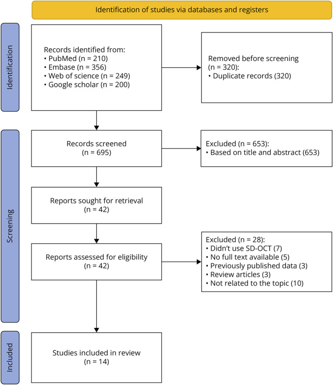
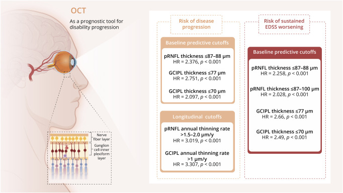
Results: Our study included 14 longitudinal studies that met our inclusion criteria, 13 studies were included in our quantitative analysis, with a total of 3,683 participants. Baseline peripapillary retinal nerve fiber layer (pRNFL) thickness below 88 μm was significantly associated with increased risk of future disease progression and physical worsening measured by Expanded Disability Status Scale progression (HR = 2.376, p < 0.001; HR = 2.258, p < 0.001, respectively). The same was noted for ganglion cell-inner plexiform layer (GCIPL) thickness below 77 μm (HR = 2.751, p < 0.001 and HR = 2.66, p < 0.001, respectively). In addition, annualized rates of pRNFL thinning above 1.5 μm/y and GCIPL thinning above 1 μm/y also significantly predicted disease worsening (HR = 3.019, p = 0.005 and HR = 3.535, p < 0.001, respectively).
Discussion: OCT-derived retinal layer thresholds, specifically a pRNFL thickness of ≤88 μm and a GCIPL thickness of ≤77 μm, are significantly associated with an increased risk of future MS disability progression. Furthermore, annual thinning rates of pRNFL >1.5 μm/y and GCIPL >1 μm/y demonstrate greater predictive power and are more clinically relevant for identifying individuals at high risk of both physical and cognitive disability progression outcomes. Further research is needed to standardize OCT thresholds and improve clinical use in treatment planning.
期刊介绍:
Neurology Neuroimmunology & Neuroinflammation is an official journal of the American Academy of Neurology. Neurology: Neuroimmunology & Neuroinflammation will be the premier peer-reviewed journal in neuroimmunology and neuroinflammation. This journal publishes rigorously peer-reviewed open-access reports of original research and in-depth reviews of topics in neuroimmunology & neuroinflammation, affecting the full range of neurologic diseases including (but not limited to) Alzheimer's disease, Parkinson's disease, ALS, tauopathy, and stroke; multiple sclerosis and NMO; inflammatory peripheral nerve and muscle disease, Guillain-Barré and myasthenia gravis; nervous system infection; paraneoplastic syndromes, noninfectious encephalitides and other antibody-mediated disorders; and psychiatric and neurodevelopmental disorders. Clinical trials, instructive case reports, and small case series will also be featured.
 求助内容:
求助内容: 应助结果提醒方式:


