{"title":"Contrast-enhanced mammography in high-dense breasts: a narrative review.","authors":"Daniele Ugo Tari, Marika Santarsiere, Davide Raffaele De Lucia, Rosalinda Santonastaso","doi":"10.21037/tbcr-24-64","DOIUrl":null,"url":null,"abstract":"<p><strong>Background and objective: </strong>Contrast-enhanced mammography (CEM) combines iodine-based contrast agents with dual-energy imaging to enhance the detection of breast cancer, especially in women with dense breast tissue. Dense breasts, which obscure conventional mammography results, present unique diagnostic challenges and an elevated risk of cancer. This review explores the role of CEM in improving diagnostic accuracy in high-density breasts, its comparative performance with other imaging modalities, and its potential implications for clinical practice.</p><p><strong>Methods: </strong>We conducted a narrative analysis on the use of CEM in dense breast tissue by searching PubMed, Web of Science (WoS), and Google Scholar between September and November 2024. Keywords, rather than MeSH terms, were utilized to refine the search, focusing on terms appearing in article titles. Sixty-six articles were identified, and duplicates or non-eligible studies were excluded, resulting in a final selection of 21 articles published between 2013 and 2024. Additional references were identified through snowballing to contextualize findings.</p><p><strong>Key content and findings: </strong>CEM demonstrates high sensitivity (89-97.7%) and specificity (50-89%) in detecting malignancies within dense breasts, offering comparable diagnostic accuracy to magnetic resonance imaging (MRI) but with better accessibility and lower cost. Unlike traditional mammography, CEM enhances visibility through functional imaging of contrast uptake, improving detection of small or occult lesions. It also aids in pre-surgical planning by assessing tumor size and multiplicity with greater precision. However, CEM is not without limitations, including radiation exposure and variability in equipment standards. Comparative analyses suggest CEM bridges the gap between conventional mammography and advanced techniques like MRI, particularly in resource-constrained settings.</p><p><strong>Conclusions: </strong>CEM represents a significant advancement in breast cancer detection, addressing limitations posed by dense breast tissue. Its diagnostic accuracy, cost-effectiveness, and patient accessibility position it as a valuable tool in personalized screening strategies. Further standardization and integration into clinical workflows could expand its role in routine breast cancer management.</p>","PeriodicalId":101427,"journal":{"name":"Translational breast cancer research : a journal focusing on translational research in breast cancer","volume":"6 ","pages":"15"},"PeriodicalIF":1.4000,"publicationDate":"2025-03-10","publicationTypes":"Journal Article","fieldsOfStudy":null,"isOpenAccess":false,"openAccessPdf":"https://www.ncbi.nlm.nih.gov/pmc/articles/PMC12104952/pdf/","citationCount":"0","resultStr":null,"platform":"Semanticscholar","paperid":null,"PeriodicalName":"Translational breast cancer research : a journal focusing on translational research in breast cancer","FirstCategoryId":"1085","ListUrlMain":"https://doi.org/10.21037/tbcr-24-64","RegionNum":0,"RegionCategory":null,"ArticlePicture":[],"TitleCN":null,"AbstractTextCN":null,"PMCID":null,"EPubDate":"2025/1/1 0:00:00","PubModel":"eCollection","JCR":"","JCRName":"","Score":null,"Total":0}
引用次数: 0
Abstract
Background and objective: Contrast-enhanced mammography (CEM) combines iodine-based contrast agents with dual-energy imaging to enhance the detection of breast cancer, especially in women with dense breast tissue. Dense breasts, which obscure conventional mammography results, present unique diagnostic challenges and an elevated risk of cancer. This review explores the role of CEM in improving diagnostic accuracy in high-density breasts, its comparative performance with other imaging modalities, and its potential implications for clinical practice.
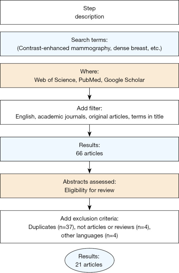
Methods: We conducted a narrative analysis on the use of CEM in dense breast tissue by searching PubMed, Web of Science (WoS), and Google Scholar between September and November 2024. Keywords, rather than MeSH terms, were utilized to refine the search, focusing on terms appearing in article titles. Sixty-six articles were identified, and duplicates or non-eligible studies were excluded, resulting in a final selection of 21 articles published between 2013 and 2024. Additional references were identified through snowballing to contextualize findings.
Key content and findings: CEM demonstrates high sensitivity (89-97.7%) and specificity (50-89%) in detecting malignancies within dense breasts, offering comparable diagnostic accuracy to magnetic resonance imaging (MRI) but with better accessibility and lower cost. Unlike traditional mammography, CEM enhances visibility through functional imaging of contrast uptake, improving detection of small or occult lesions. It also aids in pre-surgical planning by assessing tumor size and multiplicity with greater precision. However, CEM is not without limitations, including radiation exposure and variability in equipment standards. Comparative analyses suggest CEM bridges the gap between conventional mammography and advanced techniques like MRI, particularly in resource-constrained settings.
Conclusions: CEM represents a significant advancement in breast cancer detection, addressing limitations posed by dense breast tissue. Its diagnostic accuracy, cost-effectiveness, and patient accessibility position it as a valuable tool in personalized screening strategies. Further standardization and integration into clinical workflows could expand its role in routine breast cancer management.

 求助内容:
求助内容: 应助结果提醒方式:


