Elahe Hosseini, Seyyed Ali Hosseini, Stijn Servaes, Brandon Hall, Pedro Rosa-Neto, Ali-Reza Moradi, Ajay Kumar, Mir Mohsen Pedram, Sanjeev Chawla
{"title":"Transforming 3D MRI to 2D Feature Maps Using Pre-Trained Models for Diagnosis of Attention Deficit Hyperactivity Disorder.","authors":"Elahe Hosseini, Seyyed Ali Hosseini, Stijn Servaes, Brandon Hall, Pedro Rosa-Neto, Ali-Reza Moradi, Ajay Kumar, Mir Mohsen Pedram, Sanjeev Chawla","doi":"10.3390/tomography11050056","DOIUrl":null,"url":null,"abstract":"<p><p><b>Background:</b> According to the World Health Organization (WHO), approximately 5% of children and 2.5% of adults suffer from attention deficit hyperactivity disorder (ADHD). This disorder can have significant negative consequences on people's lives, particularly children. In recent years, methods based on artificial intelligence and neuroimaging techniques, such as MRI, have made significant progress, paving the way for development of more reliable diagnostic tools. In this proof of concept study, our aim was to investigate the potential utility of neuroimaging data and clinical information in combination with a deep learning-based analytical approach, more precisely, a novel feature extraction technique for the diagnosis of ADHD with high accuracy. <b>Methods:</b> Leveraging the ADHD200 dataset, which encompasses demographic information and anatomical MRI scans collected from a diverse ADHD population, our study focused on developing modern deep learning-based diagnostic models. The data preprocessing employed a pre-trained Visual Geometry Group16 (VGG16) network to extract two-dimensional (2D) feature maps from three-dimensional (3D) anatomical MRI data to reduce computational complexity and enhance diagnostic power. The inclusion of personal attributes, such as age, gender, intelligence quotient, and handedness, strengthens the diagnostic models. Four deep-learning architectures-convolutional neural network 2D (CNN2D), CNN1D, long short-term memory (LSTM), and gated recurrent units (GRU)-were employed for analysis of the MRI data, with and without the inclusion of clinical characteristics. <b>Results:</b> A 10-fold cross-validation test revealed that the LSTM model, which incorporated both MRI data and personal attributes, had the best diagnostic performance among all tested models in the diagnosis of ADHD with an accuracy of 0.86 and area under the receiver operating characteristic (ROC) curve (AUC) score of 0.90. <b>Conclusions:</b> Our findings demonstrate that the proposed approach of extracting 2D features from 3D MRI images and integrating these features with clinical characteristics may be useful in the diagnosis of ADHD with high accuracy.</p>","PeriodicalId":51330,"journal":{"name":"Tomography","volume":"11 5","pages":""},"PeriodicalIF":2.2000,"publicationDate":"2025-05-13","publicationTypes":"Journal Article","fieldsOfStudy":null,"isOpenAccess":false,"openAccessPdf":"https://www.ncbi.nlm.nih.gov/pmc/articles/PMC12115681/pdf/","citationCount":"0","resultStr":null,"platform":"Semanticscholar","paperid":null,"PeriodicalName":"Tomography","FirstCategoryId":"3","ListUrlMain":"https://doi.org/10.3390/tomography11050056","RegionNum":4,"RegionCategory":"医学","ArticlePicture":[],"TitleCN":null,"AbstractTextCN":null,"PMCID":null,"EPubDate":"","PubModel":"","JCR":"Q2","JCRName":"RADIOLOGY, NUCLEAR MEDICINE & MEDICAL IMAGING","Score":null,"Total":0}
引用次数: 0
Abstract
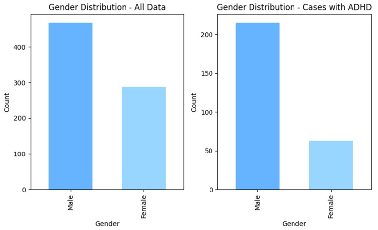
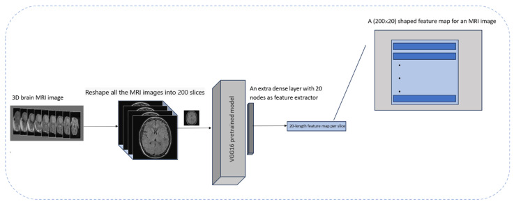
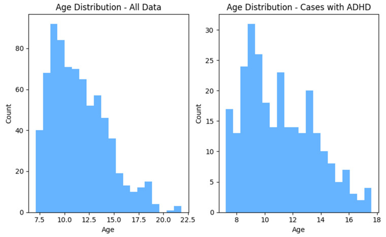
Background: According to the World Health Organization (WHO), approximately 5% of children and 2.5% of adults suffer from attention deficit hyperactivity disorder (ADHD). This disorder can have significant negative consequences on people's lives, particularly children. In recent years, methods based on artificial intelligence and neuroimaging techniques, such as MRI, have made significant progress, paving the way for development of more reliable diagnostic tools. In this proof of concept study, our aim was to investigate the potential utility of neuroimaging data and clinical information in combination with a deep learning-based analytical approach, more precisely, a novel feature extraction technique for the diagnosis of ADHD with high accuracy. Methods: Leveraging the ADHD200 dataset, which encompasses demographic information and anatomical MRI scans collected from a diverse ADHD population, our study focused on developing modern deep learning-based diagnostic models. The data preprocessing employed a pre-trained Visual Geometry Group16 (VGG16) network to extract two-dimensional (2D) feature maps from three-dimensional (3D) anatomical MRI data to reduce computational complexity and enhance diagnostic power. The inclusion of personal attributes, such as age, gender, intelligence quotient, and handedness, strengthens the diagnostic models. Four deep-learning architectures-convolutional neural network 2D (CNN2D), CNN1D, long short-term memory (LSTM), and gated recurrent units (GRU)-were employed for analysis of the MRI data, with and without the inclusion of clinical characteristics. Results: A 10-fold cross-validation test revealed that the LSTM model, which incorporated both MRI data and personal attributes, had the best diagnostic performance among all tested models in the diagnosis of ADHD with an accuracy of 0.86 and area under the receiver operating characteristic (ROC) curve (AUC) score of 0.90. Conclusions: Our findings demonstrate that the proposed approach of extracting 2D features from 3D MRI images and integrating these features with clinical characteristics may be useful in the diagnosis of ADHD with high accuracy.
TomographyMedicine-Radiology, Nuclear Medicine and Imaging
CiteScore
2.70
自引率
10.50%
发文量
222
期刊介绍:
TomographyTM publishes basic (technical and pre-clinical) and clinical scientific articles which involve the advancement of imaging technologies. Tomography encompasses studies that use single or multiple imaging modalities including for example CT, US, PET, SPECT, MR and hyperpolarization technologies, as well as optical modalities (i.e. bioluminescence, photoacoustic, endomicroscopy, fiber optic imaging and optical computed tomography) in basic sciences, engineering, preclinical and clinical medicine.
Tomography also welcomes studies involving exploration and refinement of contrast mechanisms and image-derived metrics within and across modalities toward the development of novel imaging probes for image-based feedback and intervention. The use of imaging in biology and medicine provides unparalleled opportunities to noninvasively interrogate tissues to obtain real-time dynamic and quantitative information required for diagnosis and response to interventions and to follow evolving pathological conditions. As multi-modal studies and the complexities of imaging technologies themselves are ever increasing to provide advanced information to scientists and clinicians.
Tomography provides a unique publication venue allowing investigators the opportunity to more precisely communicate integrated findings related to the diverse and heterogeneous features associated with underlying anatomical, physiological, functional, metabolic and molecular genetic activities of normal and diseased tissue. Thus Tomography publishes peer-reviewed articles which involve the broad use of imaging of any tissue and disease type including both preclinical and clinical investigations. In addition, hardware/software along with chemical and molecular probe advances are welcome as they are deemed to significantly contribute towards the long-term goal of improving the overall impact of imaging on scientific and clinical discovery.

 求助内容:
求助内容: 应助结果提醒方式:


