Yuxiao Gao, Yang Jiang, Yanhong Peng, Fujiang Yuan, Xinyue Zhang, Jianfeng Wang
{"title":"Medical Image Segmentation: A Comprehensive Review of Deep Learning-Based Methods.","authors":"Yuxiao Gao, Yang Jiang, Yanhong Peng, Fujiang Yuan, Xinyue Zhang, Jianfeng Wang","doi":"10.3390/tomography11050052","DOIUrl":null,"url":null,"abstract":"<p><p>Medical image segmentation is a critical application of computer vision in the analysis of medical images. Its primary objective is to isolate regions of interest in medical images from the background, thereby assisting clinicians in accurately identifying lesions, their sizes, locations, and their relationships with surrounding tissues. However, compared to natural images, medical images present unique challenges, such as low resolution, poor contrast, inconsistency, and scattered target regions. Furthermore, the accuracy and stability of segmentation results are subject to more stringent requirements. In recent years, with the widespread application of Convolutional Neural Networks (CNNs) in computer vision, deep learning-based methods for medical image segmentation have become a focal point of research. This paper categorizes, reviews, and summarizes the current representative methods and research status in the field of medical image segmentation. A comparative analysis of relevant experiments is presented, along with an introduction to commonly used public datasets, performance evaluation metrics, and loss functions in medical image segmentation. Finally, potential future research directions and development trends in this field are predicted and analyzed.</p>","PeriodicalId":51330,"journal":{"name":"Tomography","volume":"11 5","pages":""},"PeriodicalIF":2.2000,"publicationDate":"2025-04-30","publicationTypes":"Journal Article","fieldsOfStudy":null,"isOpenAccess":false,"openAccessPdf":"https://www.ncbi.nlm.nih.gov/pmc/articles/PMC12115501/pdf/","citationCount":"0","resultStr":null,"platform":"Semanticscholar","paperid":null,"PeriodicalName":"Tomography","FirstCategoryId":"3","ListUrlMain":"https://doi.org/10.3390/tomography11050052","RegionNum":4,"RegionCategory":"医学","ArticlePicture":[],"TitleCN":null,"AbstractTextCN":null,"PMCID":null,"EPubDate":"","PubModel":"","JCR":"Q2","JCRName":"RADIOLOGY, NUCLEAR MEDICINE & MEDICAL IMAGING","Score":null,"Total":0}
引用次数: 0
Abstract
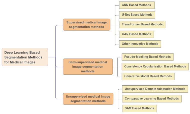
Medical image segmentation is a critical application of computer vision in the analysis of medical images. Its primary objective is to isolate regions of interest in medical images from the background, thereby assisting clinicians in accurately identifying lesions, their sizes, locations, and their relationships with surrounding tissues. However, compared to natural images, medical images present unique challenges, such as low resolution, poor contrast, inconsistency, and scattered target regions. Furthermore, the accuracy and stability of segmentation results are subject to more stringent requirements. In recent years, with the widespread application of Convolutional Neural Networks (CNNs) in computer vision, deep learning-based methods for medical image segmentation have become a focal point of research. This paper categorizes, reviews, and summarizes the current representative methods and research status in the field of medical image segmentation. A comparative analysis of relevant experiments is presented, along with an introduction to commonly used public datasets, performance evaluation metrics, and loss functions in medical image segmentation. Finally, potential future research directions and development trends in this field are predicted and analyzed.
TomographyMedicine-Radiology, Nuclear Medicine and Imaging
CiteScore
2.70
自引率
10.50%
发文量
222
期刊介绍:
TomographyTM publishes basic (technical and pre-clinical) and clinical scientific articles which involve the advancement of imaging technologies. Tomography encompasses studies that use single or multiple imaging modalities including for example CT, US, PET, SPECT, MR and hyperpolarization technologies, as well as optical modalities (i.e. bioluminescence, photoacoustic, endomicroscopy, fiber optic imaging and optical computed tomography) in basic sciences, engineering, preclinical and clinical medicine.
Tomography also welcomes studies involving exploration and refinement of contrast mechanisms and image-derived metrics within and across modalities toward the development of novel imaging probes for image-based feedback and intervention. The use of imaging in biology and medicine provides unparalleled opportunities to noninvasively interrogate tissues to obtain real-time dynamic and quantitative information required for diagnosis and response to interventions and to follow evolving pathological conditions. As multi-modal studies and the complexities of imaging technologies themselves are ever increasing to provide advanced information to scientists and clinicians.
Tomography provides a unique publication venue allowing investigators the opportunity to more precisely communicate integrated findings related to the diverse and heterogeneous features associated with underlying anatomical, physiological, functional, metabolic and molecular genetic activities of normal and diseased tissue. Thus Tomography publishes peer-reviewed articles which involve the broad use of imaging of any tissue and disease type including both preclinical and clinical investigations. In addition, hardware/software along with chemical and molecular probe advances are welcome as they are deemed to significantly contribute towards the long-term goal of improving the overall impact of imaging on scientific and clinical discovery.


 求助内容:
求助内容: 应助结果提醒方式:


