Birth-Related Subdural Hemorrhage in Asymptomatic Newborns: Magnetic Resonance Imaging Prevalence and Evolution of Intracranial and Intraspinal Localization.
IF 2.2 4区 医学Q2 RADIOLOGY, NUCLEAR MEDICINE & MEDICAL IMAGING
{"title":"Birth-Related Subdural Hemorrhage in Asymptomatic Newborns: Magnetic Resonance Imaging Prevalence and Evolution of Intracranial and Intraspinal Localization.","authors":"Davide Turilli, Leandra Piscopo, Alberto Dessì, Claudia Pinna, Liala Mirella Fattacciu, Emma Solinas, Ilaria Conti, Stefania Tamburrini, Giacomo Sica, Michele Klain, Salvatore Masala, Mariano Scaglione","doi":"10.3390/tomography11050058","DOIUrl":null,"url":null,"abstract":"<p><p><b>Background</b>: Neonatal birth-related intracranial subdural hemorrhages (SDHs) represent a form of bleeding inside the skull that occurs in newborns. This condition includes the extravasation of blood both in the encephalic parenchyma and in the extra-axial spaces. Recent studies have shown that SDH and particularly post-traumatic birth-related hemorrhages represent a frequent occurrence, but they are often asymptomatic. The gold standard for the diagnosis and follow-up of patients with SDH is multiparametric Magnetic Resonance Imaging. The aim of this study is to describe our experience by reporting several cases of SDH with different distribution and Central Nervous System involvement by the MRI of this pathology in infants up to 30 days of age. <b>Methods</b>: We analyzed the age and sex of the patients included in this study, the localization of SDH in different CNS areas, and their frequency using distribution plots and pie charts. <b>Results</b>: About the analysis of the SDH locations in the 32 patients, the most common location was the cerebellum (31/32, 96.9%), followed by parietal and occipital lobes (19/32, 59.4%; 18/32, 56.2%, respectively), falx cerebri (11/32, 34.4%), tentorium cerebelli (10/32, 31.2%), temporal lobes (6/32, 18.7%), and finally cervical and dorsal spine in the same patients (4/32, 12.5%). According to SDH locations, the patients were divided into supratentorial, infratentorial, both, and Spinal Canal. <b>Conclusions</b>: Our study confirmed the literature data regarding the neonatal birth-related SDH high frequency, but also allowed us to focus our attention on the rarest spinal SDH localizations with the same benign evolution.</p>","PeriodicalId":51330,"journal":{"name":"Tomography","volume":"11 5","pages":""},"PeriodicalIF":2.2000,"publicationDate":"2025-05-20","publicationTypes":"Journal Article","fieldsOfStudy":null,"isOpenAccess":false,"openAccessPdf":"https://www.ncbi.nlm.nih.gov/pmc/articles/PMC12115422/pdf/","citationCount":"0","resultStr":null,"platform":"Semanticscholar","paperid":null,"PeriodicalName":"Tomography","FirstCategoryId":"3","ListUrlMain":"https://doi.org/10.3390/tomography11050058","RegionNum":4,"RegionCategory":"医学","ArticlePicture":[],"TitleCN":null,"AbstractTextCN":null,"PMCID":null,"EPubDate":"","PubModel":"","JCR":"Q2","JCRName":"RADIOLOGY, NUCLEAR MEDICINE & MEDICAL IMAGING","Score":null,"Total":0}
引用次数: 0
Abstract
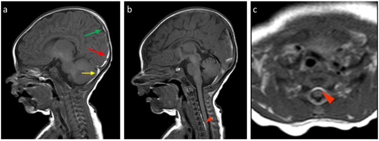
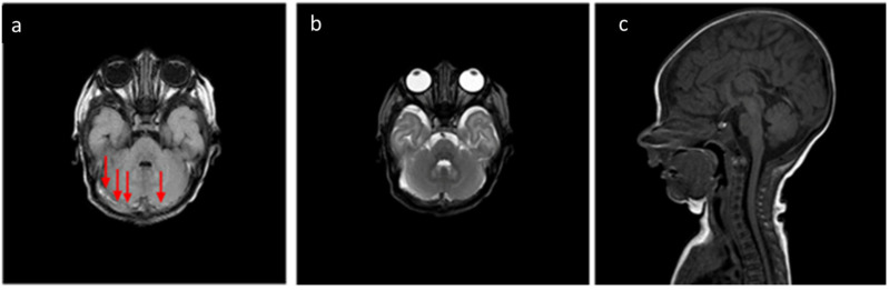
Background: Neonatal birth-related intracranial subdural hemorrhages (SDHs) represent a form of bleeding inside the skull that occurs in newborns. This condition includes the extravasation of blood both in the encephalic parenchyma and in the extra-axial spaces. Recent studies have shown that SDH and particularly post-traumatic birth-related hemorrhages represent a frequent occurrence, but they are often asymptomatic. The gold standard for the diagnosis and follow-up of patients with SDH is multiparametric Magnetic Resonance Imaging. The aim of this study is to describe our experience by reporting several cases of SDH with different distribution and Central Nervous System involvement by the MRI of this pathology in infants up to 30 days of age. Methods: We analyzed the age and sex of the patients included in this study, the localization of SDH in different CNS areas, and their frequency using distribution plots and pie charts. Results: About the analysis of the SDH locations in the 32 patients, the most common location was the cerebellum (31/32, 96.9%), followed by parietal and occipital lobes (19/32, 59.4%; 18/32, 56.2%, respectively), falx cerebri (11/32, 34.4%), tentorium cerebelli (10/32, 31.2%), temporal lobes (6/32, 18.7%), and finally cervical and dorsal spine in the same patients (4/32, 12.5%). According to SDH locations, the patients were divided into supratentorial, infratentorial, both, and Spinal Canal. Conclusions: Our study confirmed the literature data regarding the neonatal birth-related SDH high frequency, but also allowed us to focus our attention on the rarest spinal SDH localizations with the same benign evolution.
TomographyMedicine-Radiology, Nuclear Medicine and Imaging
CiteScore
2.70
自引率
10.50%
发文量
222
期刊介绍:
TomographyTM publishes basic (technical and pre-clinical) and clinical scientific articles which involve the advancement of imaging technologies. Tomography encompasses studies that use single or multiple imaging modalities including for example CT, US, PET, SPECT, MR and hyperpolarization technologies, as well as optical modalities (i.e. bioluminescence, photoacoustic, endomicroscopy, fiber optic imaging and optical computed tomography) in basic sciences, engineering, preclinical and clinical medicine.
Tomography also welcomes studies involving exploration and refinement of contrast mechanisms and image-derived metrics within and across modalities toward the development of novel imaging probes for image-based feedback and intervention. The use of imaging in biology and medicine provides unparalleled opportunities to noninvasively interrogate tissues to obtain real-time dynamic and quantitative information required for diagnosis and response to interventions and to follow evolving pathological conditions. As multi-modal studies and the complexities of imaging technologies themselves are ever increasing to provide advanced information to scientists and clinicians.
Tomography provides a unique publication venue allowing investigators the opportunity to more precisely communicate integrated findings related to the diverse and heterogeneous features associated with underlying anatomical, physiological, functional, metabolic and molecular genetic activities of normal and diseased tissue. Thus Tomography publishes peer-reviewed articles which involve the broad use of imaging of any tissue and disease type including both preclinical and clinical investigations. In addition, hardware/software along with chemical and molecular probe advances are welcome as they are deemed to significantly contribute towards the long-term goal of improving the overall impact of imaging on scientific and clinical discovery.

 求助内容:
求助内容: 应助结果提醒方式:


