David Kästner, Holger Hartmann, Robert Freudenberg, Marc Pretze, Claudia Brogsitter, Michael K Schultz, Jörg Kotzerke, Enrico Michler
{"title":"Gamma camera imaging characteristics of <sup>203/212</sup>Pb as a theragnostic pair for targeted alpha therapy: a feasibility study.","authors":"David Kästner, Holger Hartmann, Robert Freudenberg, Marc Pretze, Claudia Brogsitter, Michael K Schultz, Jörg Kotzerke, Enrico Michler","doi":"10.1186/s40658-025-00763-2","DOIUrl":null,"url":null,"abstract":"<p><strong>Background: </strong><sup>203</sup>Pb and <sup>212</sup>Pb show promise as theragnostic agents for targeted alpha therapy (TAT) because two chemically identical isotopes can be used for diagnostic imaging and treatment. In the <sup>212</sup>Pb decay chain, in addition to alpha and beta particles, a large number of photons are emitted, those with an energy of 239 keV and the characteristic X-rays of <sup>212</sup>Pb could be used for imaging. <sup>203</sup>Pb decays by photon emission with an energy of 279 keV, which appears suitable for gamma camera imaging. The aim of this study was to investigate suitable imaging protocols and to characterize the scintigraphic imaging properties and their implications for the clinical feasibility as theragnostic isotopes.</p><p><strong>Methods: </strong>Planar and SPECT/CT images were obtained with medium- and high-energy collimators on a Siemens Symbia Intevo 6 using a NEMA image quality phantom in various phantom setups and another body-shaped phantom with several inserts. Different energy windows were investigated and measurements were evaluated in terms of sensitivity, count rate performance, spatial resolution, contrast recovery, lesion detectability, and image quantification.</p><p><strong>Results: </strong>Evaluation of image quality showed superior imaging characteristics for <sup>203</sup>Pb compared to <sup>212</sup>Pb regarding spatial resolution, contrast recovery, image noise, and quantification accuracy. Both medium- and high- energy collimators were suitable for <sup>203</sup>Pb imaging, with the medium energy collimators showed slightly better imaging properties. Images obtained with the HE collimators in the 79 keV energy window showed the best visual image quality for <sup>212</sup>Pb. Due to high-energy photon emissions from <sup>212</sup>Pb daughter nuclides (e.g., 2.6 MeV from <sup>208</sup>Tl), dead time related count losses occurred even at low activities (20% count loss at 20 MBq for MELP collimators).</p><p><strong>Conclusions: </strong>According to our results and first-in-human imaging studies, SPECT/CT imaging with the <sup>203/212</sup>Pb theragnostic pair is clinically feasible. <sup>203</sup>Pb is an appropriate imaging surrogate to investigate pharmacokinetics and perform predictive dosimetry. The less favorable imaging characteristics of <sup>212</sup>Pb make image quantification and post-treatment dosimetry challenging and require further research.</p>","PeriodicalId":11559,"journal":{"name":"EJNMMI Physics","volume":"12 1","pages":"50"},"PeriodicalIF":3.2000,"publicationDate":"2025-05-27","publicationTypes":"Journal Article","fieldsOfStudy":null,"isOpenAccess":false,"openAccessPdf":"https://www.ncbi.nlm.nih.gov/pmc/articles/PMC12106261/pdf/","citationCount":"0","resultStr":null,"platform":"Semanticscholar","paperid":null,"PeriodicalName":"EJNMMI Physics","FirstCategoryId":"3","ListUrlMain":"https://doi.org/10.1186/s40658-025-00763-2","RegionNum":2,"RegionCategory":"医学","ArticlePicture":[],"TitleCN":null,"AbstractTextCN":null,"PMCID":null,"EPubDate":"","PubModel":"","JCR":"Q2","JCRName":"RADIOLOGY, NUCLEAR MEDICINE & MEDICAL IMAGING","Score":null,"Total":0}
引用次数: 0
Abstract
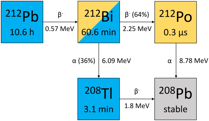
Background: 203Pb and 212Pb show promise as theragnostic agents for targeted alpha therapy (TAT) because two chemically identical isotopes can be used for diagnostic imaging and treatment. In the 212Pb decay chain, in addition to alpha and beta particles, a large number of photons are emitted, those with an energy of 239 keV and the characteristic X-rays of 212Pb could be used for imaging. 203Pb decays by photon emission with an energy of 279 keV, which appears suitable for gamma camera imaging. The aim of this study was to investigate suitable imaging protocols and to characterize the scintigraphic imaging properties and their implications for the clinical feasibility as theragnostic isotopes.
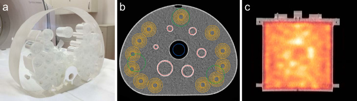
Methods: Planar and SPECT/CT images were obtained with medium- and high-energy collimators on a Siemens Symbia Intevo 6 using a NEMA image quality phantom in various phantom setups and another body-shaped phantom with several inserts. Different energy windows were investigated and measurements were evaluated in terms of sensitivity, count rate performance, spatial resolution, contrast recovery, lesion detectability, and image quantification.
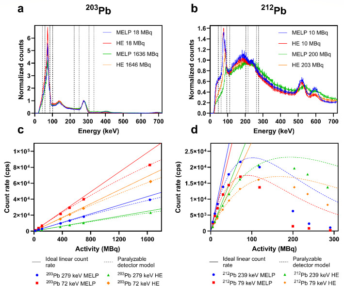
Results: Evaluation of image quality showed superior imaging characteristics for 203Pb compared to 212Pb regarding spatial resolution, contrast recovery, image noise, and quantification accuracy. Both medium- and high- energy collimators were suitable for 203Pb imaging, with the medium energy collimators showed slightly better imaging properties. Images obtained with the HE collimators in the 79 keV energy window showed the best visual image quality for 212Pb. Due to high-energy photon emissions from 212Pb daughter nuclides (e.g., 2.6 MeV from 208Tl), dead time related count losses occurred even at low activities (20% count loss at 20 MBq for MELP collimators).
Conclusions: According to our results and first-in-human imaging studies, SPECT/CT imaging with the 203/212Pb theragnostic pair is clinically feasible. 203Pb is an appropriate imaging surrogate to investigate pharmacokinetics and perform predictive dosimetry. The less favorable imaging characteristics of 212Pb make image quantification and post-treatment dosimetry challenging and require further research.
期刊介绍:
EJNMMI Physics is an international platform for scientists, users and adopters of nuclear medicine with a particular interest in physics matters. As a companion journal to the European Journal of Nuclear Medicine and Molecular Imaging, this journal has a multi-disciplinary approach and welcomes original materials and studies with a focus on applied physics and mathematics as well as imaging systems engineering and prototyping in nuclear medicine. This includes physics-driven approaches or algorithms supported by physics that foster early clinical adoption of nuclear medicine imaging and therapy.

 求助内容:
求助内容: 应助结果提醒方式:


