Anne Seewald, Jingxiao Zhong, Macarena Siri, Peter Fratzl and Emeline Raguin
{"title":"Three-dimensional imaging of vasculature and forming quail femur using cryo-correlative light and electron microscopy (cryo-CLEM)","authors":"Anne Seewald, Jingxiao Zhong, Macarena Siri, Peter Fratzl and Emeline Raguin","doi":"10.1039/D5FD00022J","DOIUrl":null,"url":null,"abstract":"<p >Bone mineralization during embryonic development requires the transport and deposition of an enormous amount of mineral precursors. In avian embryos, blood vessels play a dual role in this context: facilitating the demineralization of the eggshell to supply calcium and other minerals on the one hand, and mediating their deposition into the developing skeleton on the other. Understanding the interface between blood vessels and the surrounding tissues is therefore crucial for unraveling the mechanisms underlying biomineralization. However, visualizing this interface poses significant challenges and requires imaging methods that preserve the ultrastructure in a close-to-native state. Here we present a detailed methodology for a cryogenic correlative light and electron microscopy (cryo-CLEM) workflow to investigate the transport of mineral precursors in blood vessels of the femur of quail embryos during bone development. To achieve this, we use a fluorophore-conjugated antibody to label endothelial cells, which form the inner lining of blood vessels and which mediate exchanges between the bloodstream and developing tissues. This approach enables precise localization of blood vessels through fluorescence microscopy, which is subsequently correlated with 3D high-resolution electron microscopy using Focused Ion Beam-Scanning Electron Microscopy (FIB-SEM). This methodology allows imaging of a sufficient volume to observe both the lumen of the blood vessels and the surrounding matrix, providing deeper insights into calcium transport and bone mineralization during quail embryogenesis.</p>","PeriodicalId":49075,"journal":{"name":"Faraday Discussions","volume":"261 ","pages":" 430-445"},"PeriodicalIF":3.1000,"publicationDate":"2025-03-07","publicationTypes":"Journal Article","fieldsOfStudy":null,"isOpenAccess":false,"openAccessPdf":"https://pubs.rsc.org/en/content/articlepdf/2025/fd/d5fd00022j?page=search","citationCount":"0","resultStr":null,"platform":"Semanticscholar","paperid":null,"PeriodicalName":"Faraday Discussions","FirstCategoryId":"92","ListUrlMain":"https://pubs.rsc.org/en/content/articlelanding/2025/fd/d5fd00022j","RegionNum":3,"RegionCategory":"化学","ArticlePicture":[],"TitleCN":null,"AbstractTextCN":null,"PMCID":null,"EPubDate":"","PubModel":"","JCR":"Q2","JCRName":"Chemistry","Score":null,"Total":0}
引用次数: 0
Abstract
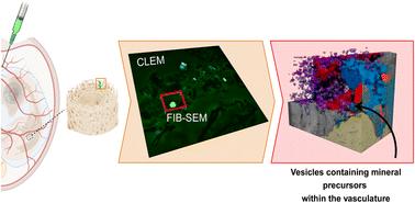
Bone mineralization during embryonic development requires the transport and deposition of an enormous amount of mineral precursors. In avian embryos, blood vessels play a dual role in this context: facilitating the demineralization of the eggshell to supply calcium and other minerals on the one hand, and mediating their deposition into the developing skeleton on the other. Understanding the interface between blood vessels and the surrounding tissues is therefore crucial for unraveling the mechanisms underlying biomineralization. However, visualizing this interface poses significant challenges and requires imaging methods that preserve the ultrastructure in a close-to-native state. Here we present a detailed methodology for a cryogenic correlative light and electron microscopy (cryo-CLEM) workflow to investigate the transport of mineral precursors in blood vessels of the femur of quail embryos during bone development. To achieve this, we use a fluorophore-conjugated antibody to label endothelial cells, which form the inner lining of blood vessels and which mediate exchanges between the bloodstream and developing tissues. This approach enables precise localization of blood vessels through fluorescence microscopy, which is subsequently correlated with 3D high-resolution electron microscopy using Focused Ion Beam-Scanning Electron Microscopy (FIB-SEM). This methodology allows imaging of a sufficient volume to observe both the lumen of the blood vessels and the surrounding matrix, providing deeper insights into calcium transport and bone mineralization during quail embryogenesis.
 求助内容:
求助内容: 应助结果提醒方式:


