{"title":"The role of FAPI PET/CT in patients with lymphoma: a systematic review.","authors":"Natale Quartuccio, Stefania Nicolosi, Sabina Pulizzi, Dante D'Oppido, Salvatore Ialuna","doi":"10.3389/fnume.2025.1589903","DOIUrl":null,"url":null,"abstract":"<p><strong>Introduction: </strong>Fluorodeoxyglucose (FDG) PET/CT is typically the reference imaging method for assessing and tracking lymphomas. However, fibroblast activation protein inhibitor (FAPI) PET is being explored as a potentially useful option, especially when Fluorodeoxyglucose (FDG) scans do not show clear results.</p><p><strong>Methods: </strong>For this systematic review, two researchers searched PubMed/MEDLINE and Cochrane CENTRAL for studies on FAPI PET/CT in lymphoma patients.</p><p><strong>Results: </strong>The literature search initially retrieved 249 articles. After removing duplicates and screening titles and abstracts, and full text, there was a final selection of 15 articles (3 original studies and 12 case reports), encompassing a total of 270 patients. The three original studies were judged to have a low risk of bias according to the QUADAS-2 criteria. The systematic review reveals that FAPI PET/CT exhibits lower diagnostic sensitivity than [<sup>18</sup>F]FDG PET/CT in lymphomas characterized by low FAP expression. Nevertheless, FAPI PET/CT retains potential as a complementary imaging modality.</p><p><strong>Discussion: </strong>[<sup>18</sup>F]FDG PET/CT remains the gold standard in lymphoma imaging, but FAPI PET/CT can potentially provide supplementary information regarding the molecular characteristics of lymphomas. FAPI PET/CT may have prognostic and therapeutic implications. In particular, it could help identify lymphoma subgroups with distinct stromal environments, potentially serving as a prognostic biomarker. Further large-scale prospective studies are warranted to validate its role in lymphoma management.</p>","PeriodicalId":73095,"journal":{"name":"Frontiers in nuclear medicine (Lausanne, Switzerland)","volume":"5 ","pages":"1589903"},"PeriodicalIF":1.4000,"publicationDate":"2025-05-09","publicationTypes":"Journal Article","fieldsOfStudy":null,"isOpenAccess":false,"openAccessPdf":"https://www.ncbi.nlm.nih.gov/pmc/articles/PMC12101064/pdf/","citationCount":"0","resultStr":null,"platform":"Semanticscholar","paperid":null,"PeriodicalName":"Frontiers in nuclear medicine (Lausanne, Switzerland)","FirstCategoryId":"1085","ListUrlMain":"https://doi.org/10.3389/fnume.2025.1589903","RegionNum":0,"RegionCategory":null,"ArticlePicture":[],"TitleCN":null,"AbstractTextCN":null,"PMCID":null,"EPubDate":"2025/1/1 0:00:00","PubModel":"eCollection","JCR":"","JCRName":"","Score":null,"Total":0}
引用次数: 0
Abstract
Introduction: Fluorodeoxyglucose (FDG) PET/CT is typically the reference imaging method for assessing and tracking lymphomas. However, fibroblast activation protein inhibitor (FAPI) PET is being explored as a potentially useful option, especially when Fluorodeoxyglucose (FDG) scans do not show clear results.
Methods: For this systematic review, two researchers searched PubMed/MEDLINE and Cochrane CENTRAL for studies on FAPI PET/CT in lymphoma patients.
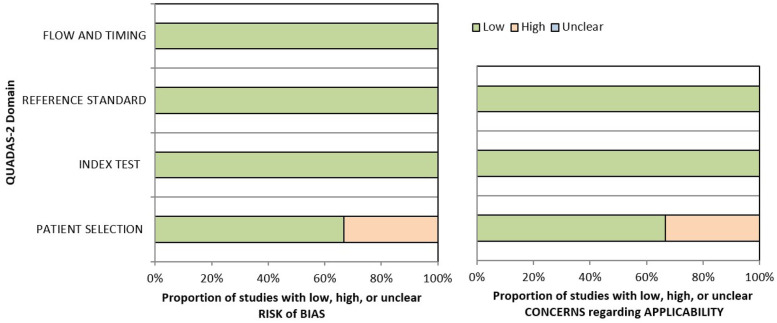
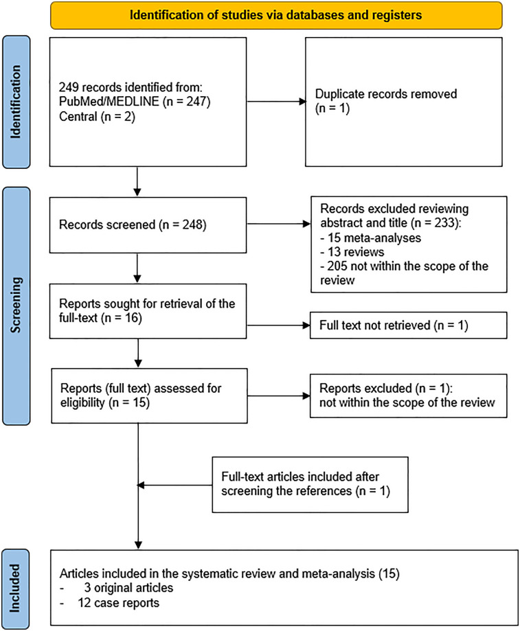
Results: The literature search initially retrieved 249 articles. After removing duplicates and screening titles and abstracts, and full text, there was a final selection of 15 articles (3 original studies and 12 case reports), encompassing a total of 270 patients. The three original studies were judged to have a low risk of bias according to the QUADAS-2 criteria. The systematic review reveals that FAPI PET/CT exhibits lower diagnostic sensitivity than [18F]FDG PET/CT in lymphomas characterized by low FAP expression. Nevertheless, FAPI PET/CT retains potential as a complementary imaging modality.
Discussion: [18F]FDG PET/CT remains the gold standard in lymphoma imaging, but FAPI PET/CT can potentially provide supplementary information regarding the molecular characteristics of lymphomas. FAPI PET/CT may have prognostic and therapeutic implications. In particular, it could help identify lymphoma subgroups with distinct stromal environments, potentially serving as a prognostic biomarker. Further large-scale prospective studies are warranted to validate its role in lymphoma management.

 求助内容:
求助内容: 应助结果提醒方式:


