Quentin A Meslier, Robert Oehrlein, Sandra J Shefelbine
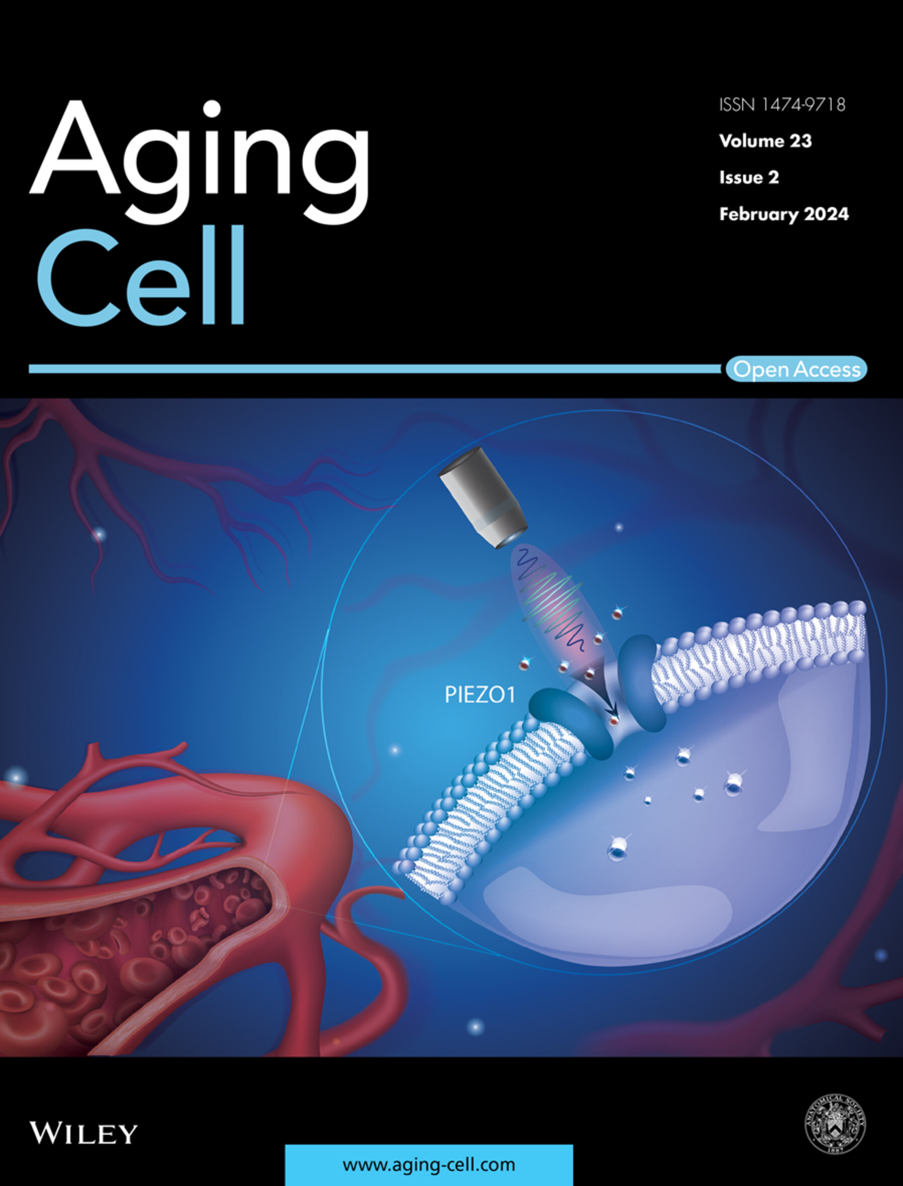
{"title":"Combined Effects of Mechanical Loading and Piezo1 Chemical Activation on 22-Months-Old Female Mouse Bone Adaptation.","authors":"Quentin A Meslier, Robert Oehrlein, Sandra J Shefelbine","doi":"10.1111/acel.70087","DOIUrl":null,"url":null,"abstract":"<p><p>With age, bones mechanosensitivity is reduced, which limits their ability to adapt to loading. The exact mechanism leading to this loss of mechanosensitvity is still unclear, making developing effective treatment challenging. Current treatments mostly focus on preventing bone mass loss (such as bisphosphonates) or promoting bone formation (such as Sclerostin inhibitors) to limit the decline of bones mass. However, treatments do not target the cause of bone mass loss which may be, in part, due to the bone's inability to initiate a normal bone mechanoadaptation response. In this work, we investigated the effects of 2 weeks of tibia loading, and Piezo1 agonist injection in vivo on 22-month-old mouse bone adaptation response. We used an optimized loading profile, which induced high fluid flow velocity and low strain magnitude in adult mouse tibia. We found that tibia loading and Yoda2 injection have an additive effect on increasing cortical bone parameters in 22-month-old mice. In vivo osteocytes calcium signaling imaging suggests that Yoda2 is able to reach osteocytes and activate Piezo1. This combination of mechanical and chemical stimulation could be a promising treatment strategy to help promote bone formation in patients who have low bone mass due to aging.</p>","PeriodicalId":119,"journal":{"name":"Aging Cell","volume":" ","pages":"e70087"},"PeriodicalIF":8.0000,"publicationDate":"2025-05-23","publicationTypes":"Journal Article","fieldsOfStudy":null,"isOpenAccess":false,"openAccessPdf":"","citationCount":"0","resultStr":null,"platform":"Semanticscholar","paperid":null,"PeriodicalName":"Aging Cell","FirstCategoryId":"99","ListUrlMain":"https://doi.org/10.1111/acel.70087","RegionNum":1,"RegionCategory":"医学","ArticlePicture":[],"TitleCN":null,"AbstractTextCN":null,"PMCID":null,"EPubDate":"","PubModel":"","JCR":"Q1","JCRName":"CELL BIOLOGY","Score":null,"Total":0}
引用次数: 0
Abstract
With age, bones mechanosensitivity is reduced, which limits their ability to adapt to loading. The exact mechanism leading to this loss of mechanosensitvity is still unclear, making developing effective treatment challenging. Current treatments mostly focus on preventing bone mass loss (such as bisphosphonates) or promoting bone formation (such as Sclerostin inhibitors) to limit the decline of bones mass. However, treatments do not target the cause of bone mass loss which may be, in part, due to the bone's inability to initiate a normal bone mechanoadaptation response. In this work, we investigated the effects of 2 weeks of tibia loading, and Piezo1 agonist injection in vivo on 22-month-old mouse bone adaptation response. We used an optimized loading profile, which induced high fluid flow velocity and low strain magnitude in adult mouse tibia. We found that tibia loading and Yoda2 injection have an additive effect on increasing cortical bone parameters in 22-month-old mice. In vivo osteocytes calcium signaling imaging suggests that Yoda2 is able to reach osteocytes and activate Piezo1. This combination of mechanical and chemical stimulation could be a promising treatment strategy to help promote bone formation in patients who have low bone mass due to aging.
Aging CellBiochemistry, Genetics and Molecular Biology-Cell Biology
自引率
2.60%
发文量
212
期刊介绍:
Aging Cell is an Open Access journal that focuses on the core aspects of the biology of aging, encompassing the entire spectrum of geroscience. The journal's content is dedicated to publishing research that uncovers the mechanisms behind the aging process and explores the connections between aging and various age-related diseases. This journal aims to provide a comprehensive understanding of the biological underpinnings of aging and its implications for human health.
The journal is widely recognized and its content is abstracted and indexed by numerous databases and services, which facilitates its accessibility and impact in the scientific community. These include:
Academic Search (EBSCO Publishing)
Academic Search Alumni Edition (EBSCO Publishing)
Academic Search Premier (EBSCO Publishing)
Biological Science Database (ProQuest)
CAS: Chemical Abstracts Service (ACS)
Embase (Elsevier)
InfoTrac (GALE Cengage)
Ingenta Select
ISI Alerting Services
Journal Citation Reports/Science Edition (Clarivate Analytics)
MEDLINE/PubMed (NLM)
Natural Science Collection (ProQuest)
PubMed Dietary Supplement Subset (NLM)
Science Citation Index Expanded (Clarivate Analytics)
SciTech Premium Collection (ProQuest)
Web of Science (Clarivate Analytics)
Being indexed in these databases ensures that the research published in Aging Cell is discoverable by researchers, clinicians, and other professionals interested in the field of aging and its associated health issues. This broad coverage helps to disseminate the journal's findings and contributes to the advancement of knowledge in geroscience.
 求助内容:
求助内容: 应助结果提醒方式:


