Mahsa Naeeni Davarani, Ali Arian Darestani, Virginia Guillen Cañas, Mohammad Hossein Harirchian, Amin Zarei, Sanaz Heydari Havadaragh, Hasan Hashemi
{"title":"Enhanced Segmentation of Active and Nonactive Multiple Sclerosis Plaques in T1 and FLAIR MRI Images Using Transformer-Based Encoders","authors":"Mahsa Naeeni Davarani, Ali Arian Darestani, Virginia Guillen Cañas, Mohammad Hossein Harirchian, Amin Zarei, Sanaz Heydari Havadaragh, Hasan Hashemi","doi":"10.1002/ima.70120","DOIUrl":null,"url":null,"abstract":"<p>Demyelinating plaques in multiple sclerosis (MS) can be visualized using magnetic resonance imaging (MRI), where accurate segmentation of active and nonactive lesions is critical for diagnosis, monitoring disease progression, and guiding treatment. Fluid-attenuated inversion recovery )FLAIR( images are widely used to detect both types of lesions, while T1-weighted images are, particularly, useful for identifying active plaques, although they are more challenging to segment due to their lower contrast and smaller lesion size. To enhance the segmentation accuracy of MS plaques, focusing on both active and non-active lesions, by utilizing TransUNet, a transformer-based neural network. The model's performance is evaluated on T1-weighted and FLAIR MRI images, with a specific focus on improving the segmentation of active plaques in T1-weighted images, which are traditionally more difficult to segment. The dataset included MRI scans from 174 patients diagnosed with MS, a significant expansion compared to previous studies. Additionally, 21 external subject test data were used to validate the model's generalizability. TransUNet was applied separately to T1-weighted and FLAIR images. Preprocessing steps included skull stripping and normalization. The model's performance was assessed using standard evaluation metrics, including Dice Coefficient, sensitivity, specificity, intersection over union (IoU), and Hausdorff distance at 95% (HD95). The study also conducted a comparative analysis between TransUNet and the widely used nnU-Net model. For FLAIR images, TransUNet achieved a sensitivity of 0.763, specificity of 0.998, IoU of 0.563, Dice coefficient of 0.712, and HD95 of 5.402 mm on the internal test set. On the external test set, it maintained a sensitivity of 0.739, specificity of 0.999, IoU of 0.551, Dice coefficient of 0.704, and HD95 of 14.630 mm. For T1-weighted images, the model showed a sensitivity of 0.494, specificity of 1.000, IoU of 0.411, Dice coefficient of 0.548, and HD95 of 22.144 mm on the internal test set. On the external test set, it improved to a sensitivity of 0.725, specificity of 0.999, IoU of 0.573, Dice coefficient of 0.693, and HD95 of 5.146 mm. Compared to nnU-Net on FLAIR images, TransUNet achieved a higher Dice coefficient (0.712 vs. 0.710) and significantly lower HD95 (5.402 vs. 28.300 mm). TransUNet significantly outperforms traditional methods, particularly in FLAIR images, demonstrating improved accuracy and boundary delineation. While T1-weighted images present challenges, the model shows potential for refinement. This study highlights the effectiveness of transformer-based architectures in medical image segmentation, suggesting TransUNet as a valuable tool for MS diagnosis and treatment monitoring.</p>","PeriodicalId":14027,"journal":{"name":"International Journal of Imaging Systems and Technology","volume":"35 3","pages":""},"PeriodicalIF":2.5000,"publicationDate":"2025-05-26","publicationTypes":"Journal Article","fieldsOfStudy":null,"isOpenAccess":false,"openAccessPdf":"https://onlinelibrary.wiley.com/doi/epdf/10.1002/ima.70120","citationCount":"0","resultStr":null,"platform":"Semanticscholar","paperid":null,"PeriodicalName":"International Journal of Imaging Systems and Technology","FirstCategoryId":"94","ListUrlMain":"https://onlinelibrary.wiley.com/doi/10.1002/ima.70120","RegionNum":4,"RegionCategory":"计算机科学","ArticlePicture":[],"TitleCN":null,"AbstractTextCN":null,"PMCID":null,"EPubDate":"","PubModel":"","JCR":"Q2","JCRName":"ENGINEERING, ELECTRICAL & ELECTRONIC","Score":null,"Total":0}
引用次数: 0
Abstract
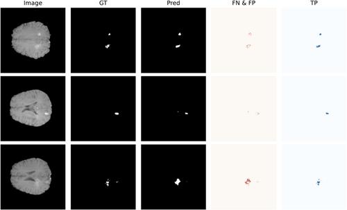
Demyelinating plaques in multiple sclerosis (MS) can be visualized using magnetic resonance imaging (MRI), where accurate segmentation of active and nonactive lesions is critical for diagnosis, monitoring disease progression, and guiding treatment. Fluid-attenuated inversion recovery )FLAIR( images are widely used to detect both types of lesions, while T1-weighted images are, particularly, useful for identifying active plaques, although they are more challenging to segment due to their lower contrast and smaller lesion size. To enhance the segmentation accuracy of MS plaques, focusing on both active and non-active lesions, by utilizing TransUNet, a transformer-based neural network. The model's performance is evaluated on T1-weighted and FLAIR MRI images, with a specific focus on improving the segmentation of active plaques in T1-weighted images, which are traditionally more difficult to segment. The dataset included MRI scans from 174 patients diagnosed with MS, a significant expansion compared to previous studies. Additionally, 21 external subject test data were used to validate the model's generalizability. TransUNet was applied separately to T1-weighted and FLAIR images. Preprocessing steps included skull stripping and normalization. The model's performance was assessed using standard evaluation metrics, including Dice Coefficient, sensitivity, specificity, intersection over union (IoU), and Hausdorff distance at 95% (HD95). The study also conducted a comparative analysis between TransUNet and the widely used nnU-Net model. For FLAIR images, TransUNet achieved a sensitivity of 0.763, specificity of 0.998, IoU of 0.563, Dice coefficient of 0.712, and HD95 of 5.402 mm on the internal test set. On the external test set, it maintained a sensitivity of 0.739, specificity of 0.999, IoU of 0.551, Dice coefficient of 0.704, and HD95 of 14.630 mm. For T1-weighted images, the model showed a sensitivity of 0.494, specificity of 1.000, IoU of 0.411, Dice coefficient of 0.548, and HD95 of 22.144 mm on the internal test set. On the external test set, it improved to a sensitivity of 0.725, specificity of 0.999, IoU of 0.573, Dice coefficient of 0.693, and HD95 of 5.146 mm. Compared to nnU-Net on FLAIR images, TransUNet achieved a higher Dice coefficient (0.712 vs. 0.710) and significantly lower HD95 (5.402 vs. 28.300 mm). TransUNet significantly outperforms traditional methods, particularly in FLAIR images, demonstrating improved accuracy and boundary delineation. While T1-weighted images present challenges, the model shows potential for refinement. This study highlights the effectiveness of transformer-based architectures in medical image segmentation, suggesting TransUNet as a valuable tool for MS diagnosis and treatment monitoring.
期刊介绍:
The International Journal of Imaging Systems and Technology (IMA) is a forum for the exchange of ideas and results relevant to imaging systems, including imaging physics and informatics. The journal covers all imaging modalities in humans and animals.
IMA accepts technically sound and scientifically rigorous research in the interdisciplinary field of imaging, including relevant algorithmic research and hardware and software development, and their applications relevant to medical research. The journal provides a platform to publish original research in structural and functional imaging.
The journal is also open to imaging studies of the human body and on animals that describe novel diagnostic imaging and analyses methods. Technical, theoretical, and clinical research in both normal and clinical populations is encouraged. Submissions describing methods, software, databases, replication studies as well as negative results are also considered.
The scope of the journal includes, but is not limited to, the following in the context of biomedical research:
Imaging and neuro-imaging modalities: structural MRI, functional MRI, PET, SPECT, CT, ultrasound, EEG, MEG, NIRS etc.;
Neuromodulation and brain stimulation techniques such as TMS and tDCS;
Software and hardware for imaging, especially related to human and animal health;
Image segmentation in normal and clinical populations;
Pattern analysis and classification using machine learning techniques;
Computational modeling and analysis;
Brain connectivity and connectomics;
Systems-level characterization of brain function;
Neural networks and neurorobotics;
Computer vision, based on human/animal physiology;
Brain-computer interface (BCI) technology;
Big data, databasing and data mining.

 求助内容:
求助内容: 应助结果提醒方式:


