Ariana Marie Martin, Andrea Paola Sanchez-Cantu, Andrea Cantu-Rodriguez, Raul Martinez-Salinas, Jesus Damian Valdez-Bocanegra
{"title":"When Giant Lymphangioma Meets Hydrops Fetalis: An Uncommon Case Report of Lower Extremity Involvement.","authors":"Ariana Marie Martin, Andrea Paola Sanchez-Cantu, Andrea Cantu-Rodriguez, Raul Martinez-Salinas, Jesus Damian Valdez-Bocanegra","doi":"10.1097/FM9.0000000000000249","DOIUrl":null,"url":null,"abstract":"<p><p>Approximately 75% of lymphangiomas manifest in the head and neck region, with limb and bone involvement observed in only 2% of cases. This case report presents a rare instance of fetal lymphangioma associated with hydrops fetalis, affecting the left lower extremity and abdomen. A 22-year-old secundigravida woman was referred to the gynecology department at 31.2 weeks of gestation. Initial fetal ultrasound revealed a live fetus with a large lymphangioma involving the left lower extremity, gluteal region, and abdomen. Additional ultrasound findings included subcutaneous edema, pericardial effusion, ascites, placentomegaly, and tricuspid regurgitation. Postnatal clinical examination confirmed the diagnosis of lymphatic malformation. Notably, only one other similar case has been reported in the English-language PubMed database. This report adds to the medical literature as a rare instance of prenatally diagnosed lymphangioma-associated hydrops fetalis.</p>","PeriodicalId":74121,"journal":{"name":"Maternal-fetal medicine (Wolters Kluwer Health, Inc.)","volume":"6 4","pages":"253-256"},"PeriodicalIF":1.7000,"publicationDate":"2024-10-11","publicationTypes":"Journal Article","fieldsOfStudy":null,"isOpenAccess":false,"openAccessPdf":"https://www.ncbi.nlm.nih.gov/pmc/articles/PMC12094381/pdf/","citationCount":"0","resultStr":null,"platform":"Semanticscholar","paperid":null,"PeriodicalName":"Maternal-fetal medicine (Wolters Kluwer Health, Inc.)","FirstCategoryId":"1085","ListUrlMain":"https://doi.org/10.1097/FM9.0000000000000249","RegionNum":0,"RegionCategory":null,"ArticlePicture":[],"TitleCN":null,"AbstractTextCN":null,"PMCID":null,"EPubDate":"2024/10/1 0:00:00","PubModel":"eCollection","JCR":"","JCRName":"","Score":null,"Total":0}
引用次数: 0
Abstract
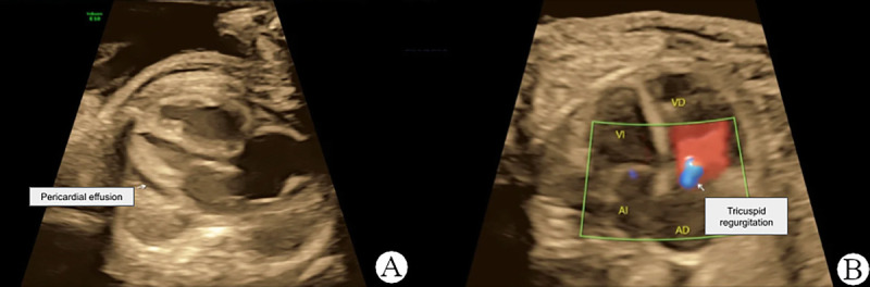
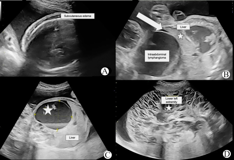
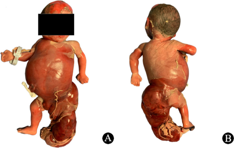
Approximately 75% of lymphangiomas manifest in the head and neck region, with limb and bone involvement observed in only 2% of cases. This case report presents a rare instance of fetal lymphangioma associated with hydrops fetalis, affecting the left lower extremity and abdomen. A 22-year-old secundigravida woman was referred to the gynecology department at 31.2 weeks of gestation. Initial fetal ultrasound revealed a live fetus with a large lymphangioma involving the left lower extremity, gluteal region, and abdomen. Additional ultrasound findings included subcutaneous edema, pericardial effusion, ascites, placentomegaly, and tricuspid regurgitation. Postnatal clinical examination confirmed the diagnosis of lymphatic malformation. Notably, only one other similar case has been reported in the English-language PubMed database. This report adds to the medical literature as a rare instance of prenatally diagnosed lymphangioma-associated hydrops fetalis.

 求助内容:
求助内容: 应助结果提醒方式:


