Rajat Vashistha, Hamed Moradi, Amanda Hammond, Kieran O'Brien, Axel Rominger, Hasan Sari, Kuangyu Shi, Viktor Vegh, David Reutens
{"title":"Non-invasive arterial input function estimation using an MRA atlas and machine learning.","authors":"Rajat Vashistha, Hamed Moradi, Amanda Hammond, Kieran O'Brien, Axel Rominger, Hasan Sari, Kuangyu Shi, Viktor Vegh, David Reutens","doi":"10.1186/s13550-025-01253-3","DOIUrl":null,"url":null,"abstract":"<p><strong>Background: </strong>Quantifying biological parameters of interest through dynamic positron emission tomography (PET) requires an arterial input function (AIF) conventionally obtained from arterial blood samples. The AIF can also be non-invasively estimated from blood pools in PET images, often identified using co-registered MRI images. Deploying methods without blood sampling or the use of MRI generally requires total body PET systems with a long axial field-of-view (LAFOV) that includes a large cardiovascular blood pool. However, the number of such systems in clinical use is currently much smaller than that of short axial field-of-view (SAFOV) scanners. We propose a data-driven approach for AIF estimation for SAFOV PET scanners, which is non-invasive and does not require MRI or blood sampling using brain PET scans. The proposed method was validated using dynamic <sup>18</sup>F-fluorodeoxyglucose [<sup>18</sup>F]FDG total body PET data from 10 subjects. A variational inference-based machine learning approach was employed to correct for peak activity. The prior was estimated using a probabilistic vascular MRI atlas, registered to each subject's PET image to identify cerebral arteries in the brain.</p><p><strong>Results: </strong>The estimated AIF using brain PET images (IDIF-Brain) was compared to that obtained using data from the descending aorta of the heart (IDIF-DA). Kinetic rate constants (K<sub>1</sub>, k<sub>2</sub>, k<sub>3</sub>) and net radiotracer influx (K<sub>i</sub>) for both cases were computed and compared. Qualitatively, the shape of IDIF-Brain matched that of IDIF-DA, capturing information on both the peak and tail of the AIF. The area under the curve (AUC) of IDIF-Brain and IDIF-DA were similar, with an average relative error of 9%. The mean Pearson correlations between kinetic parameters (K<sub>1</sub>, k<sub>2</sub>, k<sub>3</sub>) estimated with IDIF-DA and IDIF-Brain for each voxel were between 0.92 and 0.99 in all subjects, and for K<sub>i</sub>, it was above 0.97.</p><p><strong>Conclusion: </strong>This study introduces a new approach for AIF estimation in dynamic PET using brain PET images, a probabilistic vascular atlas, and machine learning techniques. The findings demonstrate the feasibility of non-invasive and subject-specific AIF estimation for SAFOV scanners.</p>","PeriodicalId":11611,"journal":{"name":"EJNMMI Research","volume":"15 1","pages":"58"},"PeriodicalIF":3.1000,"publicationDate":"2025-05-23","publicationTypes":"Journal Article","fieldsOfStudy":null,"isOpenAccess":false,"openAccessPdf":"https://www.ncbi.nlm.nih.gov/pmc/articles/PMC12102434/pdf/","citationCount":"0","resultStr":null,"platform":"Semanticscholar","paperid":null,"PeriodicalName":"EJNMMI Research","FirstCategoryId":"3","ListUrlMain":"https://doi.org/10.1186/s13550-025-01253-3","RegionNum":3,"RegionCategory":"医学","ArticlePicture":[],"TitleCN":null,"AbstractTextCN":null,"PMCID":null,"EPubDate":"","PubModel":"","JCR":"Q1","JCRName":"RADIOLOGY, NUCLEAR MEDICINE & MEDICAL IMAGING","Score":null,"Total":0}
引用次数: 0
Abstract
Background: Quantifying biological parameters of interest through dynamic positron emission tomography (PET) requires an arterial input function (AIF) conventionally obtained from arterial blood samples. The AIF can also be non-invasively estimated from blood pools in PET images, often identified using co-registered MRI images. Deploying methods without blood sampling or the use of MRI generally requires total body PET systems with a long axial field-of-view (LAFOV) that includes a large cardiovascular blood pool. However, the number of such systems in clinical use is currently much smaller than that of short axial field-of-view (SAFOV) scanners. We propose a data-driven approach for AIF estimation for SAFOV PET scanners, which is non-invasive and does not require MRI or blood sampling using brain PET scans. The proposed method was validated using dynamic 18F-fluorodeoxyglucose [18F]FDG total body PET data from 10 subjects. A variational inference-based machine learning approach was employed to correct for peak activity. The prior was estimated using a probabilistic vascular MRI atlas, registered to each subject's PET image to identify cerebral arteries in the brain.
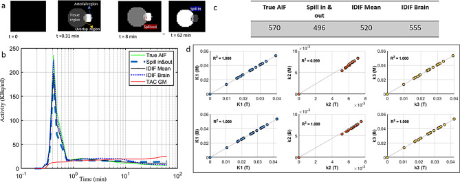
Results: The estimated AIF using brain PET images (IDIF-Brain) was compared to that obtained using data from the descending aorta of the heart (IDIF-DA). Kinetic rate constants (K1, k2, k3) and net radiotracer influx (Ki) for both cases were computed and compared. Qualitatively, the shape of IDIF-Brain matched that of IDIF-DA, capturing information on both the peak and tail of the AIF. The area under the curve (AUC) of IDIF-Brain and IDIF-DA were similar, with an average relative error of 9%. The mean Pearson correlations between kinetic parameters (K1, k2, k3) estimated with IDIF-DA and IDIF-Brain for each voxel were between 0.92 and 0.99 in all subjects, and for Ki, it was above 0.97.
Conclusion: This study introduces a new approach for AIF estimation in dynamic PET using brain PET images, a probabilistic vascular atlas, and machine learning techniques. The findings demonstrate the feasibility of non-invasive and subject-specific AIF estimation for SAFOV scanners.
EJNMMI ResearchRADIOLOGY, NUCLEAR MEDICINE & MEDICAL IMAGING&nb-
CiteScore
5.90
自引率
3.10%
发文量
72
审稿时长
13 weeks
期刊介绍:
EJNMMI Research publishes new basic, translational and clinical research in the field of nuclear medicine and molecular imaging. Regular features include original research articles, rapid communication of preliminary data on innovative research, interesting case reports, editorials, and letters to the editor. Educational articles on basic sciences, fundamental aspects and controversy related to pre-clinical and clinical research or ethical aspects of research are also welcome. Timely reviews provide updates on current applications, issues in imaging research and translational aspects of nuclear medicine and molecular imaging technologies.
The main emphasis is placed on the development of targeted imaging with radiopharmaceuticals within the broader context of molecular probes to enhance understanding and characterisation of the complex biological processes underlying disease and to develop, test and guide new treatment modalities, including radionuclide therapy.

 求助内容:
求助内容: 应助结果提醒方式:


