Serum biomarkers of collagen remodeling are associated with intestinal fibrosis and differentiate stenotic from luminal Crohn's disease patients: a pre- and post-resection longitudinal study.
Anja Poulsen, Marta Sorokina Alexdóttir, Lene Buhl Riis, Pernille Dige Ovesen, Julie Rasmussen, Mads Damsgaard Wewer, Viviane Lin, Ronja M B Lagström, Marwah Al-Sheikh, Emilie Dahl, Annedorte Ries, Martin Pehrsson, Thomai Tsapanou-Katranara, Peter-Martin Krarup, Ismail Gögenur, Florian Rieder, Johan Burisch, Joachim Høg Mortensen, Jakob Benedict Seidelin
{"title":"Serum biomarkers of collagen remodeling are associated with intestinal fibrosis and differentiate stenotic from luminal Crohn's disease patients: a pre- and post-resection longitudinal study.","authors":"Anja Poulsen, Marta Sorokina Alexdóttir, Lene Buhl Riis, Pernille Dige Ovesen, Julie Rasmussen, Mads Damsgaard Wewer, Viviane Lin, Ronja M B Lagström, Marwah Al-Sheikh, Emilie Dahl, Annedorte Ries, Martin Pehrsson, Thomai Tsapanou-Katranara, Peter-Martin Krarup, Ismail Gögenur, Florian Rieder, Johan Burisch, Joachim Høg Mortensen, Jakob Benedict Seidelin","doi":"10.1093/ecco-jcc/jjaf085","DOIUrl":null,"url":null,"abstract":"<p><strong>Background and aims: </strong>Crohn's disease (CD) is characterized by progressive intestinal transmural damage, including fibrosis and strictures, which impair quality of life and require surgical intervention. No anti-stricture therapies are available, and no accurate biomarkers have been validated allowing prediction of strictures. Collagen fragments synthesis and remodeling show potential as markers of transmural disease activity. This study aimed to evaluate serum collagen markers for their accuracy in differentiating between stenosing and luminal CD and assessing their correlation with histopathology.</p><p><strong>Methods: </strong>Sixty-two patients undergoing resection for stricturing CD and 49 with luminal CD were prospectively included. Extracellular matrix (ECM) markers were quantified using ELISA, and histological assessments of fibrosis and inflammation were performed on full-thickness tissue samples. Clinical outcomes, biomarkers, and histology were analyzed over a 12-month follow-up.</p><p><strong>Results: </strong>Extracellular matrix markers, including PRO-C6, PRO-C3, PRO-C5, C4M, and PRO-C4, distinguished stenosing from luminal CD with and the combination of PRO-C6, PRO-C3, and PRO-C5 achieved the highest discriminative power of (AUC 0.91). Significant changes in levels of the collagen biomarker were observed post-resection. Histological analysis revealed extensive intestinal fibrosis in the submucosa of the stenotic segments, which correlated with PRO-C6 levels. C4M and PRO-C4 positively correlated with neutrophils in the lamina propria. CTX-III correlated negatively with the D'Haens score and neutrophils and mononuclear cells in the lamina propria and in the epithelium.</p><p><strong>Conclusions: </strong>Collagen markers distinguished stenosing from luminal CD, and they correlated with histological fibrosis and chronic inflammation, promising for understanding ECM remodeling. This study highlights the need for extended follow-up to assess long-term stenosis-related outcomes.</p>","PeriodicalId":94074,"journal":{"name":"Journal of Crohn's & colitis","volume":" ","pages":""},"PeriodicalIF":8.7000,"publicationDate":"2025-06-04","publicationTypes":"Journal Article","fieldsOfStudy":null,"isOpenAccess":false,"openAccessPdf":"https://www.ncbi.nlm.nih.gov/pmc/articles/PMC12148806/pdf/","citationCount":"0","resultStr":null,"platform":"Semanticscholar","paperid":null,"PeriodicalName":"Journal of Crohn's & colitis","FirstCategoryId":"1085","ListUrlMain":"https://doi.org/10.1093/ecco-jcc/jjaf085","RegionNum":0,"RegionCategory":null,"ArticlePicture":[],"TitleCN":null,"AbstractTextCN":null,"PMCID":null,"EPubDate":"","PubModel":"","JCR":"","JCRName":"","Score":null,"Total":0}
引用次数: 0
Abstract
Background and aims: Crohn's disease (CD) is characterized by progressive intestinal transmural damage, including fibrosis and strictures, which impair quality of life and require surgical intervention. No anti-stricture therapies are available, and no accurate biomarkers have been validated allowing prediction of strictures. Collagen fragments synthesis and remodeling show potential as markers of transmural disease activity. This study aimed to evaluate serum collagen markers for their accuracy in differentiating between stenosing and luminal CD and assessing their correlation with histopathology.
Methods: Sixty-two patients undergoing resection for stricturing CD and 49 with luminal CD were prospectively included. Extracellular matrix (ECM) markers were quantified using ELISA, and histological assessments of fibrosis and inflammation were performed on full-thickness tissue samples. Clinical outcomes, biomarkers, and histology were analyzed over a 12-month follow-up.
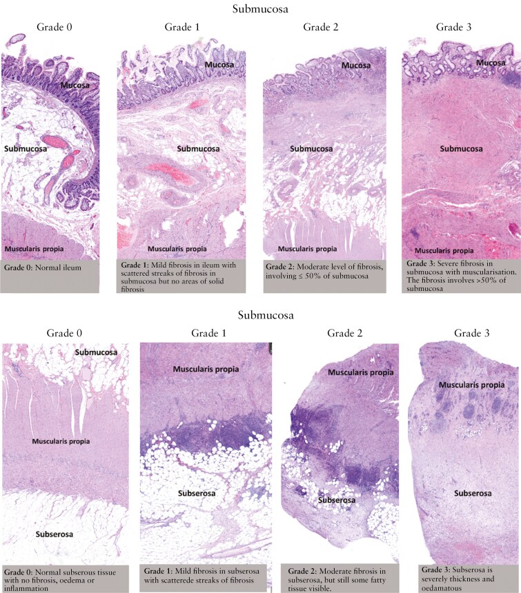
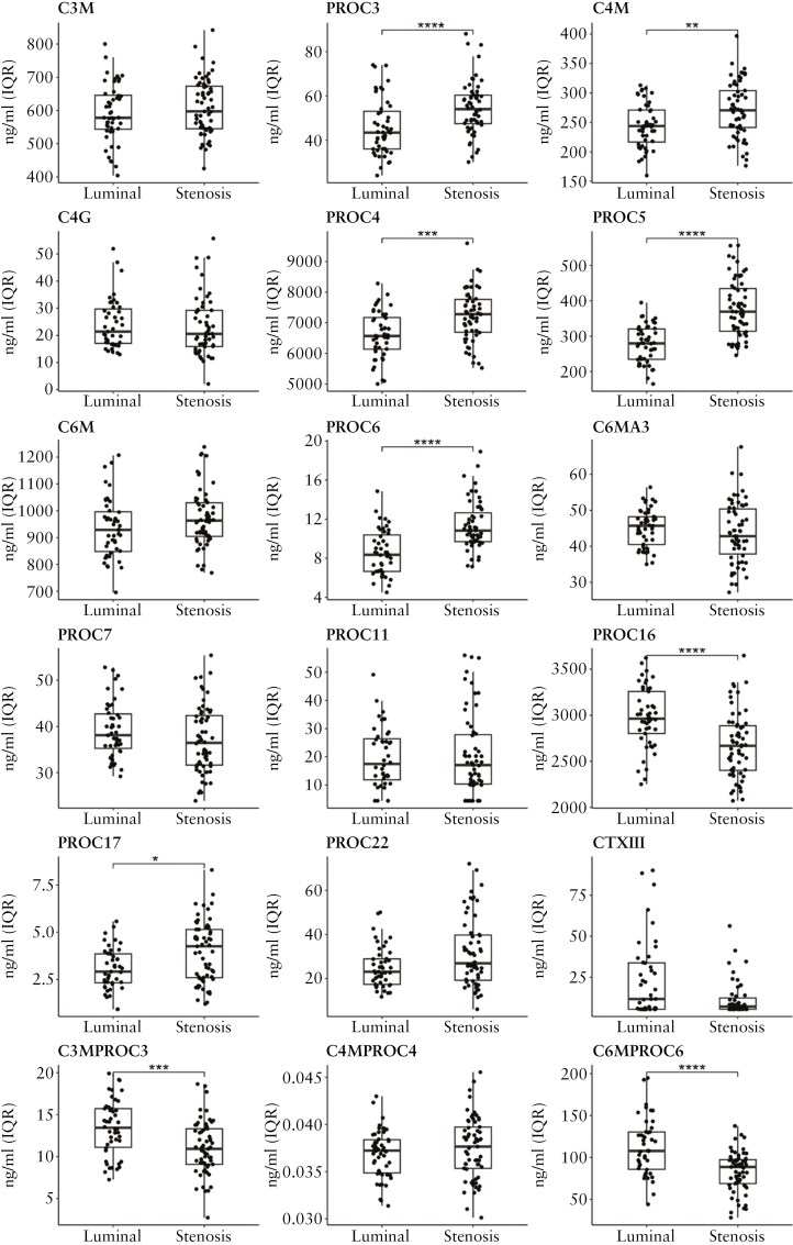
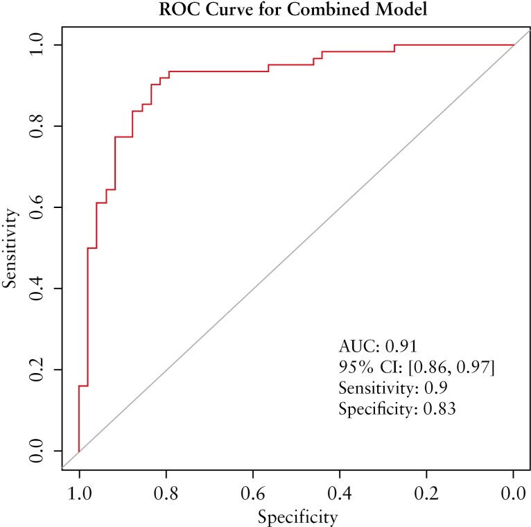
Results: Extracellular matrix markers, including PRO-C6, PRO-C3, PRO-C5, C4M, and PRO-C4, distinguished stenosing from luminal CD with and the combination of PRO-C6, PRO-C3, and PRO-C5 achieved the highest discriminative power of (AUC 0.91). Significant changes in levels of the collagen biomarker were observed post-resection. Histological analysis revealed extensive intestinal fibrosis in the submucosa of the stenotic segments, which correlated with PRO-C6 levels. C4M and PRO-C4 positively correlated with neutrophils in the lamina propria. CTX-III correlated negatively with the D'Haens score and neutrophils and mononuclear cells in the lamina propria and in the epithelium.
Conclusions: Collagen markers distinguished stenosing from luminal CD, and they correlated with histological fibrosis and chronic inflammation, promising for understanding ECM remodeling. This study highlights the need for extended follow-up to assess long-term stenosis-related outcomes.

 求助内容:
求助内容: 应助结果提醒方式:


