{"title":"A rare case of Fahr's disease with posterior circulation (basilar tip) aneurysm- pathophysiology, management, and complications.","authors":"Monirah Zeya, Vikrant Setia, Anita Jagetia","doi":"10.7461/jcen.2025.E2024.09.006","DOIUrl":null,"url":null,"abstract":"<p><p>Fahr's disease is an uncommon condition characterized by a gradual decline in cognitive, psychiatric, and motor functions, linked to idiopathic calcification in the basal ganglia, typically inherited in an autosomal dominant fashion. Acute presentation is most often as a seizure disorder; however, we present a case of an acute presentation in which the cause of the deterioration was an aneurysmal subarachnoid haemorrhage. The association between Fahr's disease and intracranial aneurysms is exceedingly rare, with only five cases documented in the literature to date. This report represents the sixth such case. Furthermore, all previously reported aneurysms were confined to the anterior circulation; thus, this is the first documented instance of Fahr's disease presenting with an aneurysm in the posterior circulation. The case here presented to the emergency service with the complaint of severe headache. Computed tomography (CT) of the head showed bilateral basal ganglia calcification and subarachnoid haemorrhage. Digital subtraction angiography (DSA) revealed a basilar tip aneurysm. The aneurysm was treated with Neqstent assisted coiling via jailing technique. Intraoperative aneurysmal haemorrhage occurred just after inserting the first coil. Heparin was reversed, blood pressure decreased and aneurysm was packed with further coils till the bleeding stopped. External ventricular drainage was performed to address subarachnoid hemorrhage (SAH) and intraventricular hemorrhage. The exact mechanisms underlying Fahr's disease are not fully understood, but it is believed to play a role in the development of aneurysms due to mineral deposits in blood vessels. For patients experiencing unexplained recurrent episodes of loss of consciousness, brain computed tomography angiography (CTA) should be performed to rule out an aneurysm, even if they have a known diagnosis of Fahr's disease, to prevent misattributing these episodes to epilepsy. Additionally, vessel wall magnetic resonance imaging (MRI) should be conducted preoperatively in cases of aneurysms linked to Fahr's disease or vasculitis to improve management planning.</p>","PeriodicalId":94072,"journal":{"name":"Journal of cerebrovascular and endovascular neurosurgery","volume":" ","pages":"276-282"},"PeriodicalIF":0.0000,"publicationDate":"2025-09-01","publicationTypes":"Journal Article","fieldsOfStudy":null,"isOpenAccess":false,"openAccessPdf":"https://www.ncbi.nlm.nih.gov/pmc/articles/PMC12488326/pdf/","citationCount":"0","resultStr":null,"platform":"Semanticscholar","paperid":null,"PeriodicalName":"Journal of cerebrovascular and endovascular neurosurgery","FirstCategoryId":"1085","ListUrlMain":"https://doi.org/10.7461/jcen.2025.E2024.09.006","RegionNum":0,"RegionCategory":null,"ArticlePicture":[],"TitleCN":null,"AbstractTextCN":null,"PMCID":null,"EPubDate":"2025/5/21 0:00:00","PubModel":"Epub","JCR":"","JCRName":"","Score":null,"Total":0}
引用次数: 0
Abstract
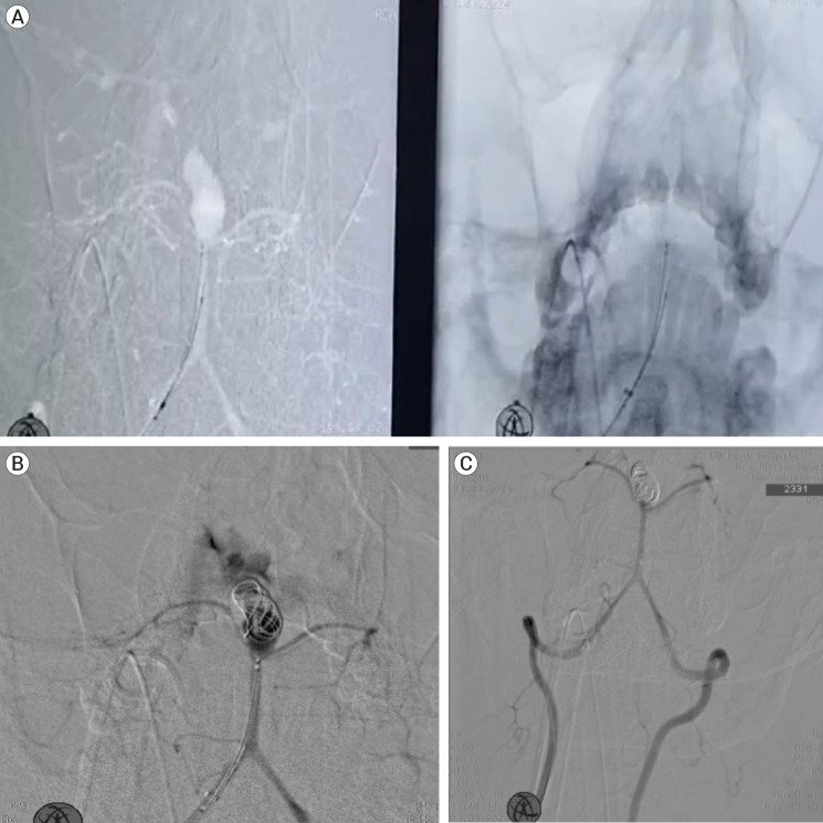
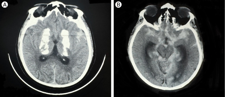
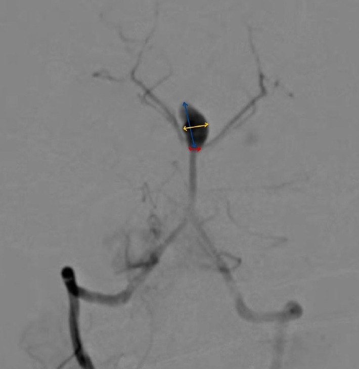
Fahr's disease is an uncommon condition characterized by a gradual decline in cognitive, psychiatric, and motor functions, linked to idiopathic calcification in the basal ganglia, typically inherited in an autosomal dominant fashion. Acute presentation is most often as a seizure disorder; however, we present a case of an acute presentation in which the cause of the deterioration was an aneurysmal subarachnoid haemorrhage. The association between Fahr's disease and intracranial aneurysms is exceedingly rare, with only five cases documented in the literature to date. This report represents the sixth such case. Furthermore, all previously reported aneurysms were confined to the anterior circulation; thus, this is the first documented instance of Fahr's disease presenting with an aneurysm in the posterior circulation. The case here presented to the emergency service with the complaint of severe headache. Computed tomography (CT) of the head showed bilateral basal ganglia calcification and subarachnoid haemorrhage. Digital subtraction angiography (DSA) revealed a basilar tip aneurysm. The aneurysm was treated with Neqstent assisted coiling via jailing technique. Intraoperative aneurysmal haemorrhage occurred just after inserting the first coil. Heparin was reversed, blood pressure decreased and aneurysm was packed with further coils till the bleeding stopped. External ventricular drainage was performed to address subarachnoid hemorrhage (SAH) and intraventricular hemorrhage. The exact mechanisms underlying Fahr's disease are not fully understood, but it is believed to play a role in the development of aneurysms due to mineral deposits in blood vessels. For patients experiencing unexplained recurrent episodes of loss of consciousness, brain computed tomography angiography (CTA) should be performed to rule out an aneurysm, even if they have a known diagnosis of Fahr's disease, to prevent misattributing these episodes to epilepsy. Additionally, vessel wall magnetic resonance imaging (MRI) should be conducted preoperatively in cases of aneurysms linked to Fahr's disease or vasculitis to improve management planning.

 求助内容:
求助内容: 应助结果提醒方式:


