Mélodie Derome, Frauke Conring, Nicole Gangl, Adamantini Hatzipanayioti, Florian Wüthrich, Maximilian Rüter, Stephanie Lefebvre, Sebastian Walther, Katharina Stegmayer
{"title":"I fear you're getting too close: neural correlates of personal space violation in paranoia.","authors":"Mélodie Derome, Frauke Conring, Nicole Gangl, Adamantini Hatzipanayioti, Florian Wüthrich, Maximilian Rüter, Stephanie Lefebvre, Sebastian Walther, Katharina Stegmayer","doi":"10.1038/s41537-025-00625-x","DOIUrl":null,"url":null,"abstract":"<p><p>Increased personal space (PS) is a clinically relevant marker for paranoia. Neuroimaging evidence suggested limbic and prefrontal circuit alterations related to threat processing and emotion regulation (i.e., amygdala, fronto-parietal cortex). We hypothesize that patients with paranoia will respond with altered activation in PS-relevant brain areas (i.e., limbic regions, fronto-parietal cortex) toward personal space intrusion. We included 79 participants with various degrees of paranoia severity; 49 patients diagnosed with schizophrenia and 30 controls. In this fMRI study, participants passively viewed pictures of facial expressions in approaching, static, or retracting motions. Violation of PS was modelled with the approaching faces condition. We used firstly a cut off to separate patients in high and low paranoia, and secondly the continuous variations of paranoia severity to understand the full picture. While participants were passively watching faces approaching them in contrast to static faces, group comparison revealed that patients with high paranoia showed hypoactivity mainly in the OFC when compared to patients with low paranoia, and hypoactivity in dlPFC and dPCC when compared to controls. Further, paranoia severity was positively associated with activation of the right hippocampus. Altered neural activity in the OFC, dlPFC, and hippocampus may well reflect the neural responses to the paranoid experience of threat and provide evidence for the hypothesized association between limbic dysfunction and paranoid threat. Modelling of paranoia severity captures variance in neural response to approaching threat, which may be previously undetected due to heterogeneity when examined at the group level.</p>","PeriodicalId":74758,"journal":{"name":"Schizophrenia (Heidelberg, Germany)","volume":"11 1","pages":"77"},"PeriodicalIF":4.1000,"publicationDate":"2025-05-21","publicationTypes":"Journal Article","fieldsOfStudy":null,"isOpenAccess":false,"openAccessPdf":"https://www.ncbi.nlm.nih.gov/pmc/articles/PMC12092760/pdf/","citationCount":"0","resultStr":null,"platform":"Semanticscholar","paperid":null,"PeriodicalName":"Schizophrenia (Heidelberg, Germany)","FirstCategoryId":"1085","ListUrlMain":"https://doi.org/10.1038/s41537-025-00625-x","RegionNum":0,"RegionCategory":null,"ArticlePicture":[],"TitleCN":null,"AbstractTextCN":null,"PMCID":null,"EPubDate":"","PubModel":"","JCR":"Q2","JCRName":"PSYCHIATRY","Score":null,"Total":0}
引用次数: 0
Abstract
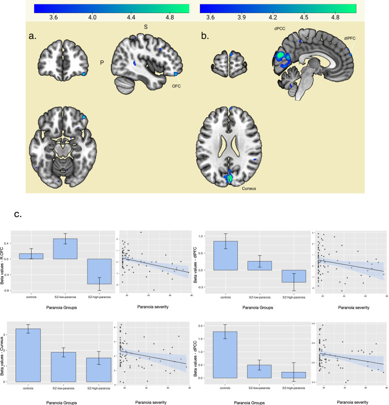
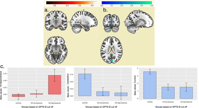
Increased personal space (PS) is a clinically relevant marker for paranoia. Neuroimaging evidence suggested limbic and prefrontal circuit alterations related to threat processing and emotion regulation (i.e., amygdala, fronto-parietal cortex). We hypothesize that patients with paranoia will respond with altered activation in PS-relevant brain areas (i.e., limbic regions, fronto-parietal cortex) toward personal space intrusion. We included 79 participants with various degrees of paranoia severity; 49 patients diagnosed with schizophrenia and 30 controls. In this fMRI study, participants passively viewed pictures of facial expressions in approaching, static, or retracting motions. Violation of PS was modelled with the approaching faces condition. We used firstly a cut off to separate patients in high and low paranoia, and secondly the continuous variations of paranoia severity to understand the full picture. While participants were passively watching faces approaching them in contrast to static faces, group comparison revealed that patients with high paranoia showed hypoactivity mainly in the OFC when compared to patients with low paranoia, and hypoactivity in dlPFC and dPCC when compared to controls. Further, paranoia severity was positively associated with activation of the right hippocampus. Altered neural activity in the OFC, dlPFC, and hippocampus may well reflect the neural responses to the paranoid experience of threat and provide evidence for the hypothesized association between limbic dysfunction and paranoid threat. Modelling of paranoia severity captures variance in neural response to approaching threat, which may be previously undetected due to heterogeneity when examined at the group level.

 求助内容:
求助内容: 应助结果提醒方式:


