Hudson McKinney, Bryan A Kirk, Anuj J Jailwala, Aaron McFarlane, Jackson L Sullivan, Raghav Agarwal, Kevin D Hiatt
{"title":"Yield of MRI in patients with spontaneous deep intracerebral hemorrhage.","authors":"Hudson McKinney, Bryan A Kirk, Anuj J Jailwala, Aaron McFarlane, Jackson L Sullivan, Raghav Agarwal, Kevin D Hiatt","doi":"10.1007/s10140-025-02348-z","DOIUrl":null,"url":null,"abstract":"<p><strong>Purpose: </strong>Hypertensive hemorrhage is the most common type of nontraumatic intracerebral hemorrhage (ICH), and it characteristically originates in deep structures, particularly the basal ganglia, internal capsules, thalami, brainstem, and cerebellum. While advanced imaging modalities like MRI can help uncover culprit lesions in cases of unexplained ICH, we hypothesized that the yield of brain MRI would be low in patients with spontaneous deep intracerebral hemorrhage.</p><p><strong>Methods: </strong>With IRB approval, we retrospectively reviewed cases of deep ICH at a single tertiary care academic center over a 5-year period and excluded cases with a known cause for hemorrhage. Patient history and demographics, initial blood pressure, and the results of the initial noncontrast head CT and subsequent imaging studies were recorded.</p><p><strong>Results: </strong>222 patients met study inclusion criteria, with a median age of 67 and 43.2% female sex. 188 patients (84.7%) had a history of hypertension, while 14 (6.3%) had a urine drug screen positive for cocaine or amphetamines during their hospital admission. The majority of hemorrhages were centered in the basal ganglia or internal capsules (116, 52.3%). Brain MRI was obtained for 120 (54.1%) of cases at a median interval of 0.97 days following the initial head CT, and of these studies, 85 (70.8%) included postcontrast imaging. Only 1 MRI study (0.8%) identified a culprit lesion adjacent to a cerebellar hematoma, which was later found to represent a pilocytic astrocytoma. 33.8% of patients overall met the modified Hong Kong Rule. Of the 77 MRIs performed in patients not meeting the modified Hong Kong Rule, 0 revealed a culprit lesion.</p><p><strong>Conclusion: </strong>Brain MRI obtained in the acute evaluation of patients with spontaneous deep intracerebral hemorrhage rarely uncovers a culprit lesion. Routine ordering of MRI in this cohort should be reconsidered, particularly in patients not meeting the modified Hong Kong Rule.</p>","PeriodicalId":11623,"journal":{"name":"Emergency Radiology","volume":" ","pages":"545-550"},"PeriodicalIF":1.3000,"publicationDate":"2025-08-01","publicationTypes":"Journal Article","fieldsOfStudy":null,"isOpenAccess":false,"openAccessPdf":"https://www.ncbi.nlm.nih.gov/pmc/articles/PMC12328518/pdf/","citationCount":"0","resultStr":null,"platform":"Semanticscholar","paperid":null,"PeriodicalName":"Emergency Radiology","FirstCategoryId":"1085","ListUrlMain":"https://doi.org/10.1007/s10140-025-02348-z","RegionNum":0,"RegionCategory":null,"ArticlePicture":[],"TitleCN":null,"AbstractTextCN":null,"PMCID":null,"EPubDate":"2025/5/20 0:00:00","PubModel":"Epub","JCR":"Q3","JCRName":"RADIOLOGY, NUCLEAR MEDICINE & MEDICAL IMAGING","Score":null,"Total":0}
引用次数: 0
Abstract
Purpose: Hypertensive hemorrhage is the most common type of nontraumatic intracerebral hemorrhage (ICH), and it characteristically originates in deep structures, particularly the basal ganglia, internal capsules, thalami, brainstem, and cerebellum. While advanced imaging modalities like MRI can help uncover culprit lesions in cases of unexplained ICH, we hypothesized that the yield of brain MRI would be low in patients with spontaneous deep intracerebral hemorrhage.
Methods: With IRB approval, we retrospectively reviewed cases of deep ICH at a single tertiary care academic center over a 5-year period and excluded cases with a known cause for hemorrhage. Patient history and demographics, initial blood pressure, and the results of the initial noncontrast head CT and subsequent imaging studies were recorded.
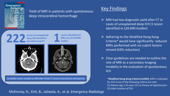
Results: 222 patients met study inclusion criteria, with a median age of 67 and 43.2% female sex. 188 patients (84.7%) had a history of hypertension, while 14 (6.3%) had a urine drug screen positive for cocaine or amphetamines during their hospital admission. The majority of hemorrhages were centered in the basal ganglia or internal capsules (116, 52.3%). Brain MRI was obtained for 120 (54.1%) of cases at a median interval of 0.97 days following the initial head CT, and of these studies, 85 (70.8%) included postcontrast imaging. Only 1 MRI study (0.8%) identified a culprit lesion adjacent to a cerebellar hematoma, which was later found to represent a pilocytic astrocytoma. 33.8% of patients overall met the modified Hong Kong Rule. Of the 77 MRIs performed in patients not meeting the modified Hong Kong Rule, 0 revealed a culprit lesion.
Conclusion: Brain MRI obtained in the acute evaluation of patients with spontaneous deep intracerebral hemorrhage rarely uncovers a culprit lesion. Routine ordering of MRI in this cohort should be reconsidered, particularly in patients not meeting the modified Hong Kong Rule.
期刊介绍:
To advance and improve the radiologic aspects of emergency careTo establish Emergency Radiology as an area of special interest in the field of diagnostic imagingTo improve methods of education in Emergency RadiologyTo provide, through formal meetings, a mechanism for presentation of scientific papers on various aspects of Emergency Radiology and continuing educationTo promote research in Emergency Radiology by clinical and basic science investigators, including residents and other traineesTo act as the resource body on Emergency Radiology for those interested in emergency patient care Members of the American Society of Emergency Radiology (ASER) receive the Emergency Radiology journal as a benefit of membership!


 求助内容:
求助内容: 应助结果提醒方式:


