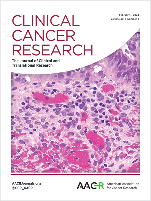
Spatial transcriptomics advances the use of canine patients in cancer research: analysis of osteosarcoma-bearing pet dogs enrolled in a clinical trial.
IF 10
1区 医学
Q1 ONCOLOGY
引用次数: 0
Abstract
PURPOSE Pet dogs spontaneously develop many of the same tumor types as humans including osteosarcoma. Recent advances in spatial transcriptomics have improved our ability to utilize formalin-fixed paraffin-embedded tissues collected at canine autopsy. These techniques allow the canine model to be investigated alongside murine models to inform human cancer research. Herein we present the first application of the GeoMx Canine Cancer Atlas to outcome-linked samples from canine patients enrolled in an osteosarcoma clinical trial. EXPERIMENTAL DESIGN A tissue microarray (TMA) of primary osteosarcoma samples was assayed using the GeoMx Digital Spatial Profiler. Samples were stratified by disease-free interval (DFI). Patients within the upper and lower tertiles were assigned to the High (n = 8) and Low DFI (n = 8) groups, respectively. Analyses included the identification of differentially expressed genes, pathway enrichment, and cell deconvolution. RESULTS Genes enriched in High DFI tumors included PTEN and CDKN1B. Low DFI tumors were enriched for NCAM1. Pathways enriched in the Low DFI group included MYC, MTORC1, and oxidative phosphorylation. High DFI tumors were enriched for interferon alpha response and allograft rejection pathways among others. Macrophages and CD8+ T cells were elevated in High DFI tumors. CONCLUSIONS Gene and pathway enrichment found to be associated with disease-free interval in this study overlap with that described in human osteosarcoma patients underscoring the value of the canine model in metastatic osteosarcoma research.空间转录组学促进了犬患者在癌症研究中的应用:对参加临床试验的患有骨肉瘤的宠物狗的分析。
目的:宠物狗会自发地发展出许多与人类相同的肿瘤类型,包括骨肉瘤。空间转录组学的最新进展提高了我们利用福尔马林固定石蜡包埋组织收集犬解剖的能力。这些技术允许犬类模型与小鼠模型一起进行研究,从而为人类癌症研究提供信息。在此,我们首次将GeoMx犬癌症图谱应用于骨肉瘤临床试验中犬患者的结果相关样本。实验设计使用GeoMx数字空间分析器对原发性骨肉瘤样本进行组织微阵列(TMA)检测。样本按无病间隔(DFI)分层。上半部分和下半部分的患者分别被分配到高(n = 8)和低DFI (n = 8)组。分析包括鉴定差异表达基因,途径富集和细胞反褶积。结果高DFI肿瘤中富集的基因包括PTEN和CDKN1B。低DFI肿瘤富集NCAM1。低DFI组中富集的通路包括MYC、MTORC1和氧化磷酸化。高DFI肿瘤富含干扰素α反应和同种异体移植排斥途径等。巨噬细胞和CD8+ T细胞在高DFI肿瘤中升高。结论本研究中发现的与无病期相关的基因和通路富集与人类骨肉瘤患者的无病期相关的基因和通路富集重叠,强调了犬模型在转移性骨肉瘤研究中的价值。
本文章由计算机程序翻译,如有差异,请以英文原文为准。
求助全文
约1分钟内获得全文
求助全文
来源期刊

Clinical Cancer Research
医学-肿瘤学
CiteScore
20.10
自引率
1.70%
发文量
1207
审稿时长
2.1 months
期刊介绍:
Clinical Cancer Research is a journal focusing on groundbreaking research in cancer, specifically in the areas where the laboratory and the clinic intersect. Our primary interest lies in clinical trials that investigate novel treatments, accompanied by research on pharmacology, molecular alterations, and biomarkers that can predict response or resistance to these treatments. Furthermore, we prioritize laboratory and animal studies that explore new drugs and targeted agents with the potential to advance to clinical trials. We also encourage research on targetable mechanisms of cancer development, progression, and metastasis.
 求助内容:
求助内容: 应助结果提醒方式:


