Francesca Baessato, Alessandro Ruzzarin, Christian Meierhofer
{"title":"Cardiovascular magnetic resonance and valvular heart diseases: a suggested protocol for congenital lesions.","authors":"Francesca Baessato, Alessandro Ruzzarin, Christian Meierhofer","doi":"10.21037/cdt-24-470","DOIUrl":null,"url":null,"abstract":"<p><p>Valvular heart diseases (VHDs) require definition of anatomy, severity, and risk stratification to best define procedural need, type of intervention and seriate follow-up. Congenital lesions are much rarer and often associated with more complex lesions. Among noninvasive imaging modalities, cardiovascular magnetic resonance (CMR) represents a fundamental tool for complete assessment and quantification of VHDs. CMR can provide wide anatomical views on cardiac and extra-cardiac structures in any plane orientation, flow and volume quantification, as well as information on ventricular remodeling and viability. In the context of valve stenosis, quantification by CMR is based primarily on direct measurement of valve orifice at maximal valve opening, although CMR data remain less reliable than standard echocardiography due to reduced temporal resolution. Definition of great vessels anatomy by CMR can allow differentiation of valvular, subvalvular or supravalvular lesions. For valve regurgitation, CMR is the gold standard for quantification of ventricular volumes and function and for direct calculation of regurgitation of the semilunar valves with through-plane phase-contrast images. Additional flow measurements can be integrated to cross-check quantitative data on great vessels flow and stroke volumes. A standardized approach is recommended in CMR studies. A minimum CMR dataset should include two-dimensional cine and phase-contrast sequences, and three-dimensional whole heart imaging. This should be applied in the clinical practice to assess VHDs, including most complex congenital lesions.</p>","PeriodicalId":9592,"journal":{"name":"Cardiovascular diagnosis and therapy","volume":"15 2","pages":"441-454"},"PeriodicalIF":2.1000,"publicationDate":"2025-04-30","publicationTypes":"Journal Article","fieldsOfStudy":null,"isOpenAccess":false,"openAccessPdf":"https://www.ncbi.nlm.nih.gov/pmc/articles/PMC12082240/pdf/","citationCount":"0","resultStr":null,"platform":"Semanticscholar","paperid":null,"PeriodicalName":"Cardiovascular diagnosis and therapy","FirstCategoryId":"3","ListUrlMain":"https://doi.org/10.21037/cdt-24-470","RegionNum":3,"RegionCategory":"医学","ArticlePicture":[],"TitleCN":null,"AbstractTextCN":null,"PMCID":null,"EPubDate":"2025/4/23 0:00:00","PubModel":"Epub","JCR":"Q3","JCRName":"CARDIAC & CARDIOVASCULAR SYSTEMS","Score":null,"Total":0}
引用次数: 0
Abstract
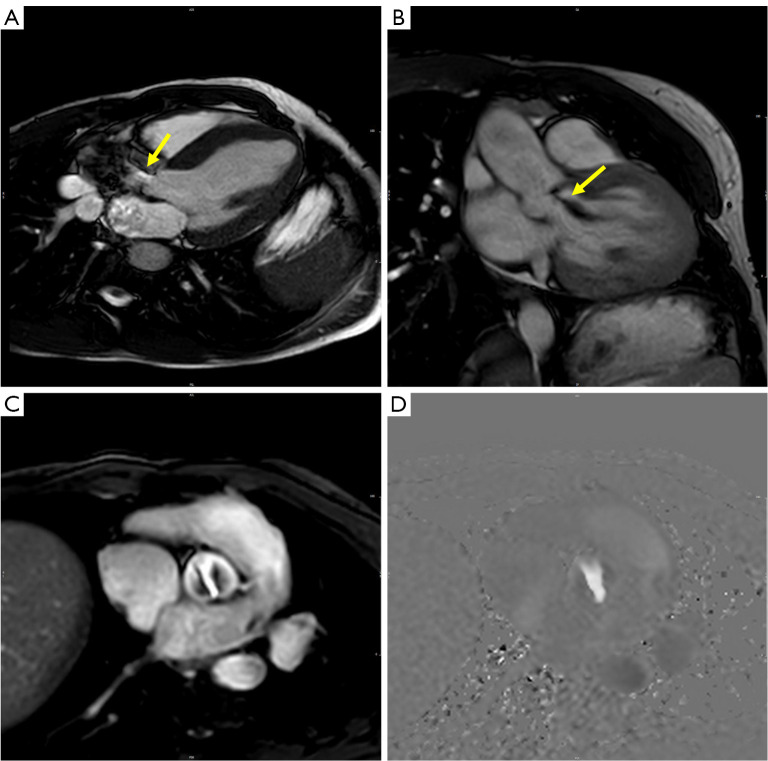
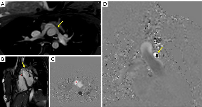
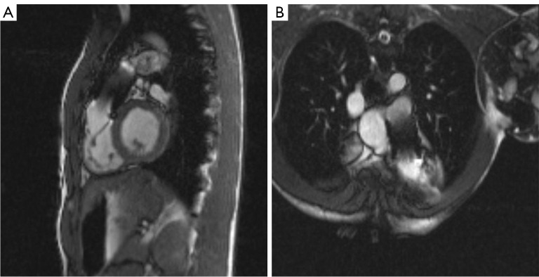
Valvular heart diseases (VHDs) require definition of anatomy, severity, and risk stratification to best define procedural need, type of intervention and seriate follow-up. Congenital lesions are much rarer and often associated with more complex lesions. Among noninvasive imaging modalities, cardiovascular magnetic resonance (CMR) represents a fundamental tool for complete assessment and quantification of VHDs. CMR can provide wide anatomical views on cardiac and extra-cardiac structures in any plane orientation, flow and volume quantification, as well as information on ventricular remodeling and viability. In the context of valve stenosis, quantification by CMR is based primarily on direct measurement of valve orifice at maximal valve opening, although CMR data remain less reliable than standard echocardiography due to reduced temporal resolution. Definition of great vessels anatomy by CMR can allow differentiation of valvular, subvalvular or supravalvular lesions. For valve regurgitation, CMR is the gold standard for quantification of ventricular volumes and function and for direct calculation of regurgitation of the semilunar valves with through-plane phase-contrast images. Additional flow measurements can be integrated to cross-check quantitative data on great vessels flow and stroke volumes. A standardized approach is recommended in CMR studies. A minimum CMR dataset should include two-dimensional cine and phase-contrast sequences, and three-dimensional whole heart imaging. This should be applied in the clinical practice to assess VHDs, including most complex congenital lesions.
期刊介绍:
The journal ''Cardiovascular Diagnosis and Therapy'' (Print ISSN: 2223-3652; Online ISSN: 2223-3660) accepts basic and clinical science submissions related to Cardiovascular Medicine and Surgery. The mission of the journal is the rapid exchange of scientific information between clinicians and scientists worldwide. To reach this goal, the journal will focus on novel media, using a web-based, digital format in addition to traditional print-version. This includes on-line submission, review, publication, and distribution. The digital format will also allow submission of extensive supporting visual material, both images and video. The website www.thecdt.org will serve as the central hub and also allow posting of comments and on-line discussion. The web-site of the journal will be linked to a number of international web-sites (e.g. www.dxy.cn), which will significantly expand the distribution of its contents.

 求助内容:
求助内容: 应助结果提醒方式:


