Natalia Konovalova, Aniket Tolpadi, Felix Liu, Zehra Akkaya, Johanna Luitjens, Felix Gassert, Paula Giesler, Rupsa Bhattacharjee, Misung Han, Emma Bahroos, Sharmila Majumdar, Valentina Pedoia
{"title":"Improving radiologist detection of meniscal abnormality on undersampled, deep learning reconstructed knee MRI.","authors":"Natalia Konovalova, Aniket Tolpadi, Felix Liu, Zehra Akkaya, Johanna Luitjens, Felix Gassert, Paula Giesler, Rupsa Bhattacharjee, Misung Han, Emma Bahroos, Sharmila Majumdar, Valentina Pedoia","doi":"10.1093/radadv/umaf015","DOIUrl":null,"url":null,"abstract":"<p><strong>Background: </strong>Accurate interpretation of meniscal anomalies on knee MRI is critical for diagnosis and treatment planning, with artificial intelligence emerging as a promising tool to support and enhance this process through automated anomaly detection.</p><p><strong>Purpose: </strong>To evaluate the impact of an artificial intelligence (AI) anomaly detection assistant on radiologists' interpretation of meniscal anomalies in undersampled, deep learning (DL)-reconstructed knee MRI and assess the relationship between reconstruction quality metrics and anomaly detection performance.</p><p><strong>Materials and methods: </strong>This retrospective study included 947 knee MRI examinations; 51 were excluded for poor image quality, leaving 896 participants (mean age, 44.7 ± 15.3 years; 472 women). Using 8-fold undersampled data, DL-based reconstructed images were generated. An object detection model was trained on original, fully sampled images and evaluated on 1 original and 14 DL-reconstructed test sets to identify meniscal lesions. Standard reconstruction metrics (normalized root mean square error, peak signal-to-noise ratio, and structural similarity index) and anomaly detection metrics (mean average precision, F1 score) were quantified and compared. Two radiologists independently reviewed a stratified sample of 50 examinations unassisted and assisted with AI-predicted anomaly boxes. McNemar's test evaluated differences in diagnostic performance; Cohen's kappa assessed interrater agreement.</p><p><strong>Results: </strong>On the original images, the anomaly detection model achieved the following: 70.53% precision, 72.17% recall, 63.09% mAP, and a 71.34% F1 score. Comparing performance among the undersampled reconstruction datasets, box-based reconstruction metrics showed better correlation with detection performance than traditional image-based metrics (mAP to box-based SSIM, <i>r</i> = 0.81, <i>P</i> < .01; mAP to image-based SSIM, <i>r</i> = 0.64, <i>P</i> = .01). In 50 participants, AI assistance improved radiologists' accuracy on reconstructed images. Sensitivity increased from 77.27% (95% CI, 65.83-85.72; 51/66) to 80.30% (95% CI, 69.16-88.11; 53/66), and specificity improved from 88.46% (95% CI, 83.73-91.95; 207/234) to 90.60% (95% CI, 86.18-93.71; 212/234) (<i>P</i> < .05).</p><p><strong>Conclusion: </strong>AI-assisted meniscal anomaly detection enhanced radiologists' interpretation of undersampled, DL-reconstructed knee MRI. Anomaly detection may serve as a complementary tool alongside other reconstruction metrics to assess the preservation of clinically important features in reconstructed images, warranting further investigation.</p>","PeriodicalId":519940,"journal":{"name":"Radiology advances","volume":"2 2","pages":"umaf015"},"PeriodicalIF":0.0000,"publicationDate":"2025-04-04","publicationTypes":"Journal Article","fieldsOfStudy":null,"isOpenAccess":false,"openAccessPdf":"https://www.ncbi.nlm.nih.gov/pmc/articles/PMC12021832/pdf/","citationCount":"0","resultStr":null,"platform":"Semanticscholar","paperid":null,"PeriodicalName":"Radiology advances","FirstCategoryId":"1085","ListUrlMain":"https://doi.org/10.1093/radadv/umaf015","RegionNum":0,"RegionCategory":null,"ArticlePicture":[],"TitleCN":null,"AbstractTextCN":null,"PMCID":null,"EPubDate":"2025/3/1 0:00:00","PubModel":"eCollection","JCR":"","JCRName":"","Score":null,"Total":0}
引用次数: 0
Abstract
Background: Accurate interpretation of meniscal anomalies on knee MRI is critical for diagnosis and treatment planning, with artificial intelligence emerging as a promising tool to support and enhance this process through automated anomaly detection.
Purpose: To evaluate the impact of an artificial intelligence (AI) anomaly detection assistant on radiologists' interpretation of meniscal anomalies in undersampled, deep learning (DL)-reconstructed knee MRI and assess the relationship between reconstruction quality metrics and anomaly detection performance.
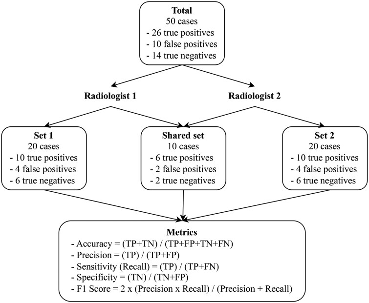
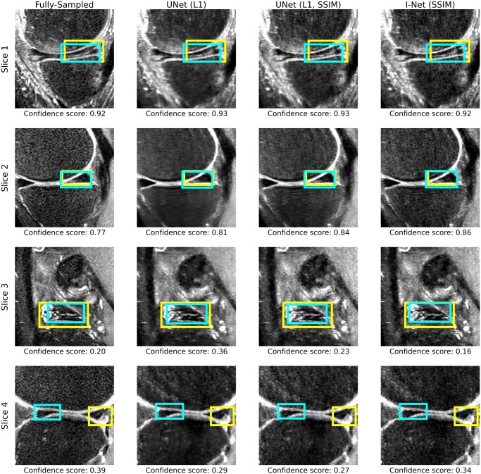
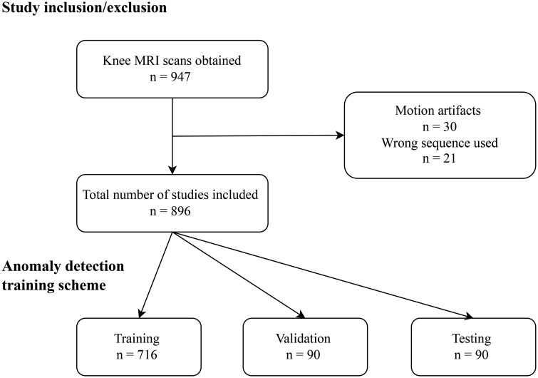
Materials and methods: This retrospective study included 947 knee MRI examinations; 51 were excluded for poor image quality, leaving 896 participants (mean age, 44.7 ± 15.3 years; 472 women). Using 8-fold undersampled data, DL-based reconstructed images were generated. An object detection model was trained on original, fully sampled images and evaluated on 1 original and 14 DL-reconstructed test sets to identify meniscal lesions. Standard reconstruction metrics (normalized root mean square error, peak signal-to-noise ratio, and structural similarity index) and anomaly detection metrics (mean average precision, F1 score) were quantified and compared. Two radiologists independently reviewed a stratified sample of 50 examinations unassisted and assisted with AI-predicted anomaly boxes. McNemar's test evaluated differences in diagnostic performance; Cohen's kappa assessed interrater agreement.
Results: On the original images, the anomaly detection model achieved the following: 70.53% precision, 72.17% recall, 63.09% mAP, and a 71.34% F1 score. Comparing performance among the undersampled reconstruction datasets, box-based reconstruction metrics showed better correlation with detection performance than traditional image-based metrics (mAP to box-based SSIM, r = 0.81, P < .01; mAP to image-based SSIM, r = 0.64, P = .01). In 50 participants, AI assistance improved radiologists' accuracy on reconstructed images. Sensitivity increased from 77.27% (95% CI, 65.83-85.72; 51/66) to 80.30% (95% CI, 69.16-88.11; 53/66), and specificity improved from 88.46% (95% CI, 83.73-91.95; 207/234) to 90.60% (95% CI, 86.18-93.71; 212/234) (P < .05).
Conclusion: AI-assisted meniscal anomaly detection enhanced radiologists' interpretation of undersampled, DL-reconstructed knee MRI. Anomaly detection may serve as a complementary tool alongside other reconstruction metrics to assess the preservation of clinically important features in reconstructed images, warranting further investigation.

 求助内容:
求助内容: 应助结果提醒方式:


