Kaan Pota, Celal Çağrı Baysal, Osman Civan, Mustafa Ürgüden
{"title":"Evaluation of the relations between foot & ankle pathologies and anatomic variations with magnetic resonance imaging of 849 study population.","authors":"Kaan Pota, Celal Çağrı Baysal, Osman Civan, Mustafa Ürgüden","doi":"10.52312/jdrs.2025.2146","DOIUrl":null,"url":null,"abstract":"<p><strong>Objectives: </strong>The aim of the study was to evaluate the relationships between common pathologies and anatomical variations in the foot & ankle using magnetic resonance imaging (MRI).</p><p><strong>Patients and methods: </strong>Between January 2016 and December 2020, a total of 849 ankle MRIs (427 right foot, 422 left foot) in 738 patients (274 males, 464 females; mean age: 43.4±14.3 years; range, 15 to 70 years) were retrospectively analyzed. Among ankle pathologies, peroneal and flexor hallucis longus (FHL) tendinopathies were evaluated. Among the anatomical variations, retromalleolar fibular groove (RMFG) shape, os peroneum, os trigonum, peroneus quartus (PQ), flexor digitorum accessorius longus (FDAL), low-lying peroneus brevis (PB) and FHL muscles were examined. The distance of the PB and FHL musculotendinous junctions (MTJs) from designated reference points was measured. Cut-off values for PB and FHL musculotendinous junction distances were determined by receiver operating characteristic (ROC) analysis. For the reliability analysis of measurements performed by two researchers, intraclass correlation coefficient (ICC) values were calculated.</p><p><strong>Results: </strong>Bilateral ankle MRIs of 111 patients were evaluated. The PB, PL, and FHL tenosynovitis were observed in 29.6%, 34.9%, and 38.8% of all ankles, respectively. The PB and PL tendon tears were found in 12.2% and 3.9%, respectively. A total of 47.1% of the RMFG shapes were concave, 36.7% were flat, 12.4% were convex, and 3.8% were irregular. The PQ, FDAL, os peroneum, and os trigonum were detected in 13.8%, 3.1%, 16.6%, and 20.5% of the ankles, respectively. The cut-off value of PB MTJ distance that would cause a PB tendon tear was 4.40 mm distal from reference point. The cut-off value of FHL MTJ distance that would cause FHL tendinopathy was 4.15 mm distal from reference point. The study had a statistically significantly high level of consistency between the experts (ICC=0.85).</p><p><strong>Conclusion: </strong>The convex and irregular shapes of the RMFG, along with the anatomical variations of the os peroneum and low-lying PB muscle, constitute risk factors for peroneal tendon pathologies. The presence of the os trigonum and low-lying FHL muscle anatomical variations predispose individuals to FHL tendinopathies. The cut-off values that could lead to PB vertical tears and FHL tendinopathy were identified for the low-lying PB and FHL muscles, respectively.</p>","PeriodicalId":73560,"journal":{"name":"Joint diseases and related surgery","volume":"36 2","pages":"394-407"},"PeriodicalIF":1.9000,"publicationDate":"2025-04-09","publicationTypes":"Journal Article","fieldsOfStudy":null,"isOpenAccess":false,"openAccessPdf":"https://www.ncbi.nlm.nih.gov/pmc/articles/PMC12086489/pdf/","citationCount":"0","resultStr":null,"platform":"Semanticscholar","paperid":null,"PeriodicalName":"Joint diseases and related surgery","FirstCategoryId":"1085","ListUrlMain":"https://doi.org/10.52312/jdrs.2025.2146","RegionNum":0,"RegionCategory":null,"ArticlePicture":[],"TitleCN":null,"AbstractTextCN":null,"PMCID":null,"EPubDate":"","PubModel":"","JCR":"Q2","JCRName":"ORTHOPEDICS","Score":null,"Total":0}
引用次数: 0
Abstract
Objectives: The aim of the study was to evaluate the relationships between common pathologies and anatomical variations in the foot & ankle using magnetic resonance imaging (MRI).
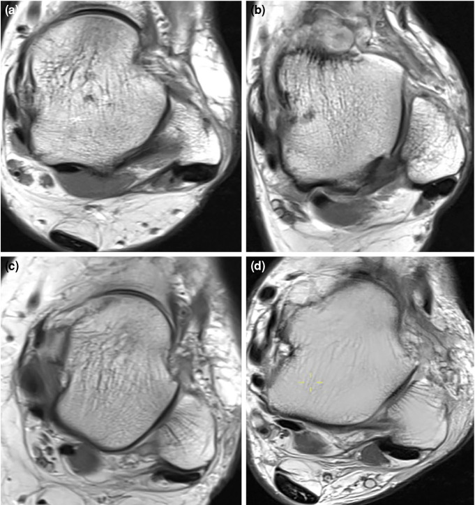
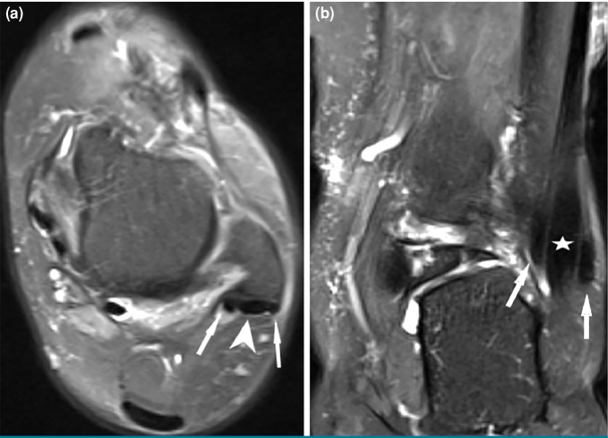
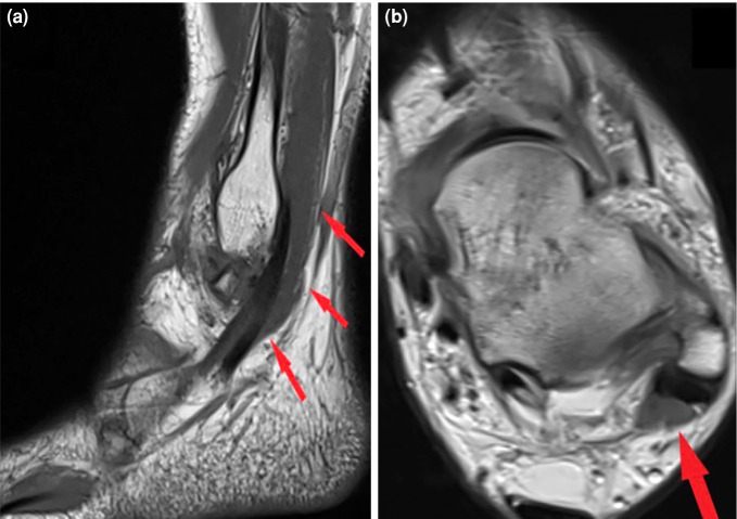
Patients and methods: Between January 2016 and December 2020, a total of 849 ankle MRIs (427 right foot, 422 left foot) in 738 patients (274 males, 464 females; mean age: 43.4±14.3 years; range, 15 to 70 years) were retrospectively analyzed. Among ankle pathologies, peroneal and flexor hallucis longus (FHL) tendinopathies were evaluated. Among the anatomical variations, retromalleolar fibular groove (RMFG) shape, os peroneum, os trigonum, peroneus quartus (PQ), flexor digitorum accessorius longus (FDAL), low-lying peroneus brevis (PB) and FHL muscles were examined. The distance of the PB and FHL musculotendinous junctions (MTJs) from designated reference points was measured. Cut-off values for PB and FHL musculotendinous junction distances were determined by receiver operating characteristic (ROC) analysis. For the reliability analysis of measurements performed by two researchers, intraclass correlation coefficient (ICC) values were calculated.
Results: Bilateral ankle MRIs of 111 patients were evaluated. The PB, PL, and FHL tenosynovitis were observed in 29.6%, 34.9%, and 38.8% of all ankles, respectively. The PB and PL tendon tears were found in 12.2% and 3.9%, respectively. A total of 47.1% of the RMFG shapes were concave, 36.7% were flat, 12.4% were convex, and 3.8% were irregular. The PQ, FDAL, os peroneum, and os trigonum were detected in 13.8%, 3.1%, 16.6%, and 20.5% of the ankles, respectively. The cut-off value of PB MTJ distance that would cause a PB tendon tear was 4.40 mm distal from reference point. The cut-off value of FHL MTJ distance that would cause FHL tendinopathy was 4.15 mm distal from reference point. The study had a statistically significantly high level of consistency between the experts (ICC=0.85).
Conclusion: The convex and irregular shapes of the RMFG, along with the anatomical variations of the os peroneum and low-lying PB muscle, constitute risk factors for peroneal tendon pathologies. The presence of the os trigonum and low-lying FHL muscle anatomical variations predispose individuals to FHL tendinopathies. The cut-off values that could lead to PB vertical tears and FHL tendinopathy were identified for the low-lying PB and FHL muscles, respectively.

 求助内容:
求助内容: 应助结果提醒方式:


