Khaled Abu Arif, Ioan Stefan Florian, Alexandru Ioan Florian, Alina Vasilica Blesneag, Enola Maer, Răzvan Mircea Cherecheș
{"title":"Assessing Acute DWI Lesions in Clinically Diagnosed TIA: Insights from a Cohort Study in Cluj, Romania.","authors":"Khaled Abu Arif, Ioan Stefan Florian, Alexandru Ioan Florian, Alina Vasilica Blesneag, Enola Maer, Răzvan Mircea Cherecheș","doi":"10.3390/tomography11040040","DOIUrl":null,"url":null,"abstract":"<p><strong>Background: </strong>The updated definition of a TIA emphasizes tissue characteristics rather than symptom duration, defining a TIA as a transient neurological episode without ischemic lesions in brain imaging, including in DWI. If imaging reveals a lesion, even in patients with transient symptoms, the event is reclassified as a minor ischemic stroke.</p><p><strong>Objective: </strong>This retrospective observational study aimed to determine the prevalence of ischemic lesions in DWI in patients with a TIA diagnosis.</p><p><strong>Results: </strong>Adults aged 18-90 years, diagnosed with a TIA by a neurologist and who underwent MRI-DWI at CMT hospital within the first week after symptom onset (May 2023-July 2024), were included. Ethical approval was obtained. Descriptive statistics summarized patient demographics, clinical features, Fazekas scale grades, and imaging findings.</p><p><strong>Conclusions: </strong>Among the 26 patients clinically diagnosed with TIAs, 7 (26.9%) exhibited ischemic lesions in DWI, reclassifying these cases as minor ischemic strokes under the updated definition. The prevalence of ischemic lesions was notably higher in patients with comorbidities such as hypertension and diabetes. These findings highlight the importance of early MRI-DWI to accurately distinguish TIAs from minor ischemic strokes. Routine urgent DWI within the first week of TIA symptoms enhances diagnosis and risk stratification and can guide targeted stroke prevention strategies, particularly when combined with the ABCD2 score.</p>","PeriodicalId":51330,"journal":{"name":"Tomography","volume":"11 4","pages":""},"PeriodicalIF":2.2000,"publicationDate":"2025-03-27","publicationTypes":"Journal Article","fieldsOfStudy":null,"isOpenAccess":false,"openAccessPdf":"https://www.ncbi.nlm.nih.gov/pmc/articles/PMC12031323/pdf/","citationCount":"0","resultStr":null,"platform":"Semanticscholar","paperid":null,"PeriodicalName":"Tomography","FirstCategoryId":"3","ListUrlMain":"https://doi.org/10.3390/tomography11040040","RegionNum":4,"RegionCategory":"医学","ArticlePicture":[],"TitleCN":null,"AbstractTextCN":null,"PMCID":null,"EPubDate":"","PubModel":"","JCR":"Q2","JCRName":"RADIOLOGY, NUCLEAR MEDICINE & MEDICAL IMAGING","Score":null,"Total":0}
引用次数: 0
Abstract
Background: The updated definition of a TIA emphasizes tissue characteristics rather than symptom duration, defining a TIA as a transient neurological episode without ischemic lesions in brain imaging, including in DWI. If imaging reveals a lesion, even in patients with transient symptoms, the event is reclassified as a minor ischemic stroke.
Objective: This retrospective observational study aimed to determine the prevalence of ischemic lesions in DWI in patients with a TIA diagnosis.
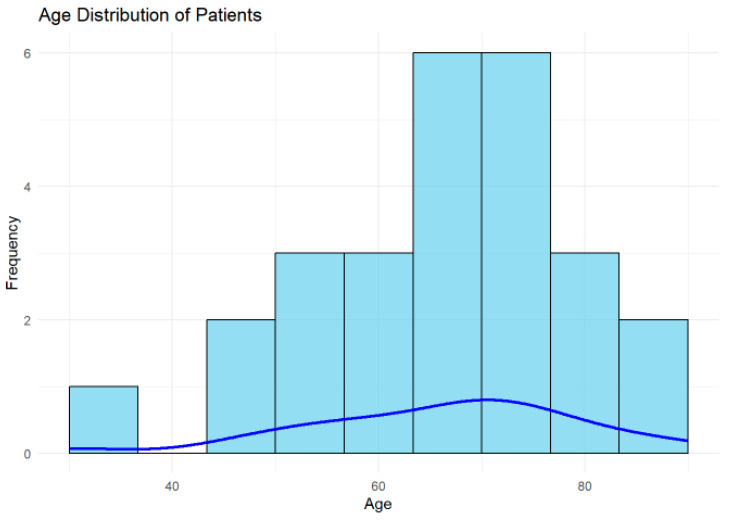
Results: Adults aged 18-90 years, diagnosed with a TIA by a neurologist and who underwent MRI-DWI at CMT hospital within the first week after symptom onset (May 2023-July 2024), were included. Ethical approval was obtained. Descriptive statistics summarized patient demographics, clinical features, Fazekas scale grades, and imaging findings.
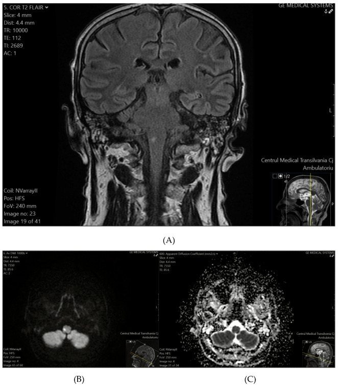
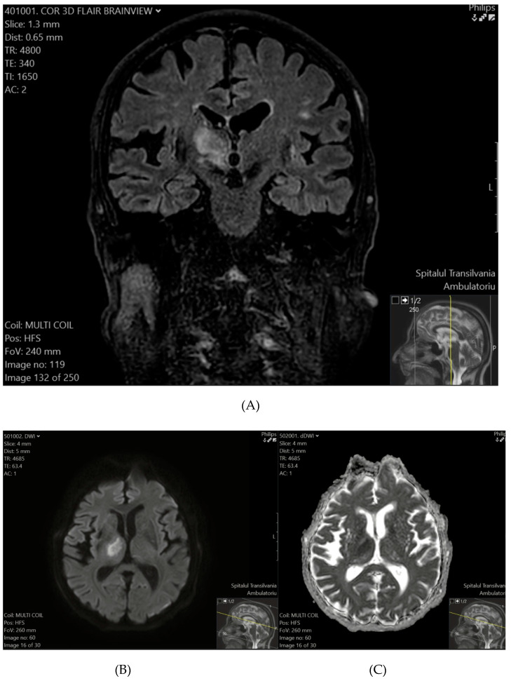
Conclusions: Among the 26 patients clinically diagnosed with TIAs, 7 (26.9%) exhibited ischemic lesions in DWI, reclassifying these cases as minor ischemic strokes under the updated definition. The prevalence of ischemic lesions was notably higher in patients with comorbidities such as hypertension and diabetes. These findings highlight the importance of early MRI-DWI to accurately distinguish TIAs from minor ischemic strokes. Routine urgent DWI within the first week of TIA symptoms enhances diagnosis and risk stratification and can guide targeted stroke prevention strategies, particularly when combined with the ABCD2 score.
TomographyMedicine-Radiology, Nuclear Medicine and Imaging
CiteScore
2.70
自引率
10.50%
发文量
222
期刊介绍:
TomographyTM publishes basic (technical and pre-clinical) and clinical scientific articles which involve the advancement of imaging technologies. Tomography encompasses studies that use single or multiple imaging modalities including for example CT, US, PET, SPECT, MR and hyperpolarization technologies, as well as optical modalities (i.e. bioluminescence, photoacoustic, endomicroscopy, fiber optic imaging and optical computed tomography) in basic sciences, engineering, preclinical and clinical medicine.
Tomography also welcomes studies involving exploration and refinement of contrast mechanisms and image-derived metrics within and across modalities toward the development of novel imaging probes for image-based feedback and intervention. The use of imaging in biology and medicine provides unparalleled opportunities to noninvasively interrogate tissues to obtain real-time dynamic and quantitative information required for diagnosis and response to interventions and to follow evolving pathological conditions. As multi-modal studies and the complexities of imaging technologies themselves are ever increasing to provide advanced information to scientists and clinicians.
Tomography provides a unique publication venue allowing investigators the opportunity to more precisely communicate integrated findings related to the diverse and heterogeneous features associated with underlying anatomical, physiological, functional, metabolic and molecular genetic activities of normal and diseased tissue. Thus Tomography publishes peer-reviewed articles which involve the broad use of imaging of any tissue and disease type including both preclinical and clinical investigations. In addition, hardware/software along with chemical and molecular probe advances are welcome as they are deemed to significantly contribute towards the long-term goal of improving the overall impact of imaging on scientific and clinical discovery.

 求助内容:
求助内容: 应助结果提醒方式:


