Ahmet Emrah Açan, Aslı Karakılıç, Mert Emre Aydın, Özgür Bulmuş, Emrah Özcan, Gülay Turan, Özge Özmen, Reşit Buğra Hüsemoğlu
{"title":"Comparison of tendon healing using local platelet-rich plasma, erythropoietin, and erythropoietin-bevacizumab in a rat Achilles tenotomy model.","authors":"Ahmet Emrah Açan, Aslı Karakılıç, Mert Emre Aydın, Özgür Bulmuş, Emrah Özcan, Gülay Turan, Özge Özmen, Reşit Buğra Hüsemoğlu","doi":"10.52312/jdrs.2025.2234","DOIUrl":null,"url":null,"abstract":"<p><strong>Objectives: </strong>This study aims to evaluate the effects of platelet-rich plasma (PRP), erythropoietin (EPO), and EPO-bevacizumab (EPO-BEVA) combination on tendon healing in a rat Achilles tenotomy model.</p><p><strong>Materials and methods: </strong>Fifty-six male Wistar albino rats (14 to 16 weeks old) were randomly assigned to control, PRP, EPO, and EPO-BEVA groups including 14 rats in each group. Bilateral Achilles tenotomy was performed under anesthesia, followed by respective treatments. Platelet-rich plasma (0.1 mL/tendon) was prepared using a Ficoll-based extraction kit. The EPO (500 U/kg) and EPO-BEVA (175 U EPO + 1.25 mg BEVA) were administered locally. Biomechanical analysis assessed maximum force, stiffness, tensile stress, and Young's modulus. Histological evaluation included Bonar scoring, collagen organization, tenocyte morphology, and vascularity. Cross-sectional area (CSA) was measured.</p><p><strong>Results: </strong>At Week 2, the EPO-BEVA group exhibited superior stiffness (14.79±6.9 N/mm) than PRP (8.64±1.5 N/mm, p=0.015) and greater tensile stress (8.2±1 MPa) than control (6.16±1.3 MPa, p=0.031). The CSA was reduced (4.79±0.8 mm<sup>2</sup>) compared to EPO (6.56±1.1 mm<sup>2</sup>, p=0.038), indicating qualitative tendon improvements. Histological analysis showed enhanced matrix organization and reduced vascularity in the EPO-BEVA group, with lower Bonar scores (5.29±1.4 vs. 9.29±1.1 in control, p=0.002). By Week 4, maximum force remained higher in EPO-BEVA (46.67±5.8 N) than control (34.84±3 N, p=0.004), with sustained Young's modulus superiority compared to EPO (3.2±1.2 MPa vs. 1.78±0.5 MPa, p=0.014), although the stiffness differences were no longer significant.</p><p><strong>Conclusion: </strong>Our study results showed that EPO-BEVA enhanced tendon healing via vascular and matrix modulation, although the lack of a BEVA-only group limits conclusions on synergy. Future studies with larger sample sizes, including BEVA monotherapy, optimized dosing strategies, and long-term evaluations are needed to better clarify these effects and refine treatment strategies in regenerative medicine.</p>","PeriodicalId":73560,"journal":{"name":"Joint diseases and related surgery","volume":"36 2","pages":"383-393"},"PeriodicalIF":1.9000,"publicationDate":"2025-04-05","publicationTypes":"Journal Article","fieldsOfStudy":null,"isOpenAccess":false,"openAccessPdf":"https://www.ncbi.nlm.nih.gov/pmc/articles/PMC12086474/pdf/","citationCount":"0","resultStr":null,"platform":"Semanticscholar","paperid":null,"PeriodicalName":"Joint diseases and related surgery","FirstCategoryId":"1085","ListUrlMain":"https://doi.org/10.52312/jdrs.2025.2234","RegionNum":0,"RegionCategory":null,"ArticlePicture":[],"TitleCN":null,"AbstractTextCN":null,"PMCID":null,"EPubDate":"","PubModel":"","JCR":"Q2","JCRName":"ORTHOPEDICS","Score":null,"Total":0}
引用次数: 0
Abstract
Objectives: This study aims to evaluate the effects of platelet-rich plasma (PRP), erythropoietin (EPO), and EPO-bevacizumab (EPO-BEVA) combination on tendon healing in a rat Achilles tenotomy model.
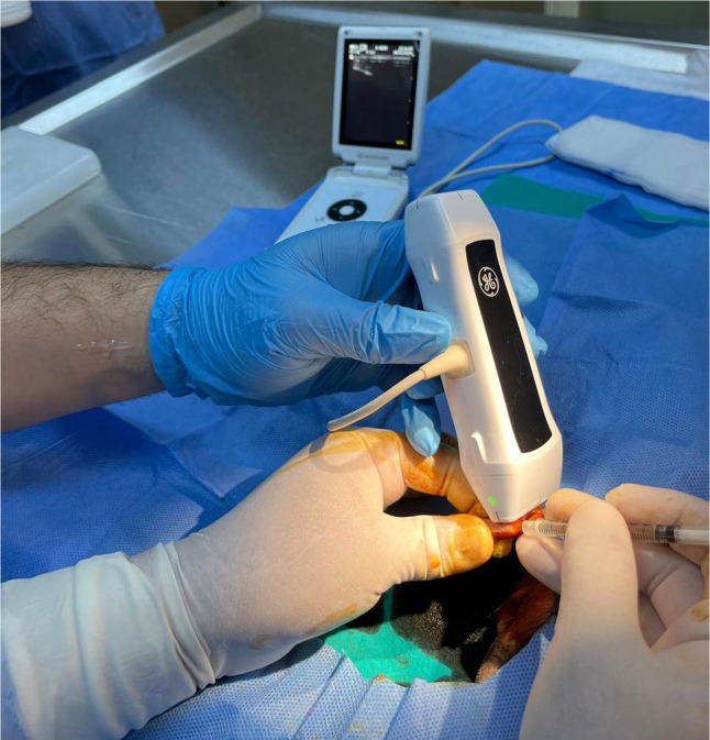
Materials and methods: Fifty-six male Wistar albino rats (14 to 16 weeks old) were randomly assigned to control, PRP, EPO, and EPO-BEVA groups including 14 rats in each group. Bilateral Achilles tenotomy was performed under anesthesia, followed by respective treatments. Platelet-rich plasma (0.1 mL/tendon) was prepared using a Ficoll-based extraction kit. The EPO (500 U/kg) and EPO-BEVA (175 U EPO + 1.25 mg BEVA) were administered locally. Biomechanical analysis assessed maximum force, stiffness, tensile stress, and Young's modulus. Histological evaluation included Bonar scoring, collagen organization, tenocyte morphology, and vascularity. Cross-sectional area (CSA) was measured.
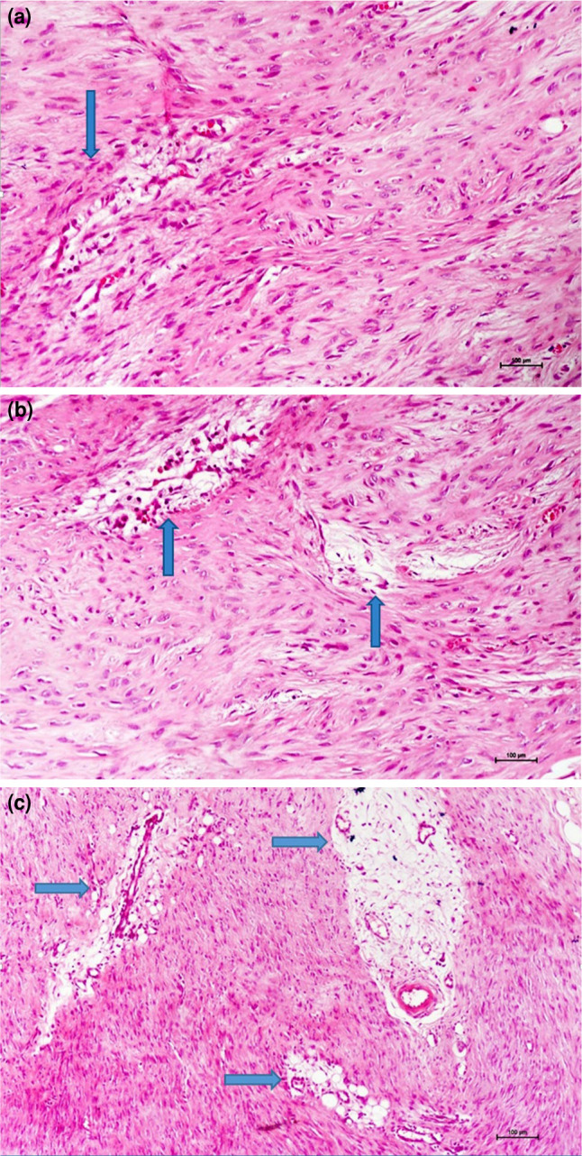
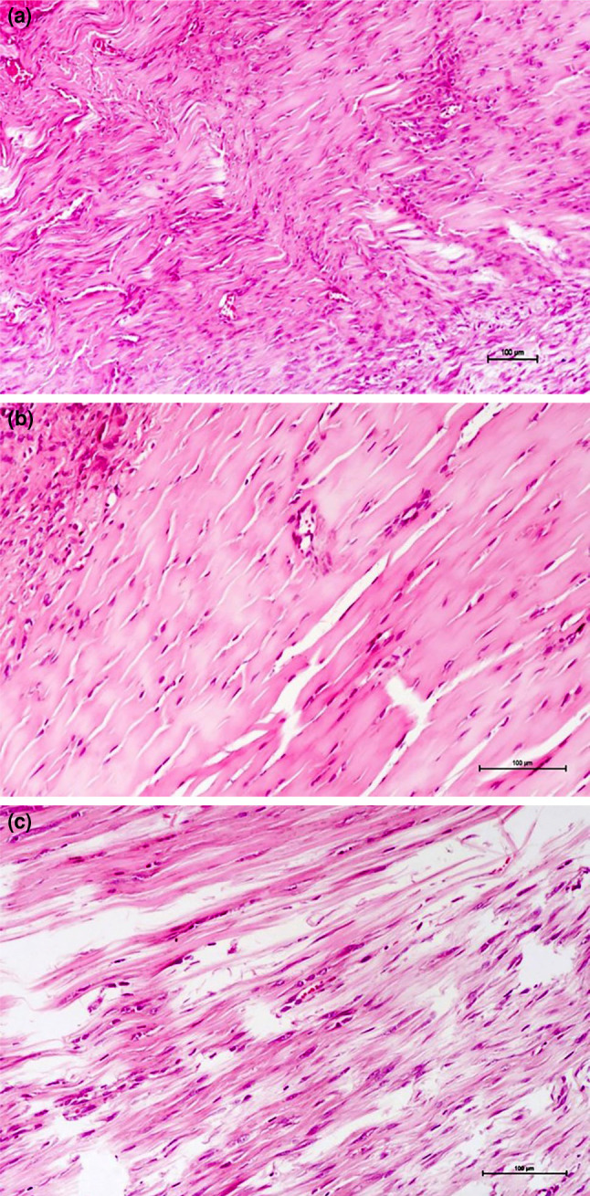
Results: At Week 2, the EPO-BEVA group exhibited superior stiffness (14.79±6.9 N/mm) than PRP (8.64±1.5 N/mm, p=0.015) and greater tensile stress (8.2±1 MPa) than control (6.16±1.3 MPa, p=0.031). The CSA was reduced (4.79±0.8 mm2) compared to EPO (6.56±1.1 mm2, p=0.038), indicating qualitative tendon improvements. Histological analysis showed enhanced matrix organization and reduced vascularity in the EPO-BEVA group, with lower Bonar scores (5.29±1.4 vs. 9.29±1.1 in control, p=0.002). By Week 4, maximum force remained higher in EPO-BEVA (46.67±5.8 N) than control (34.84±3 N, p=0.004), with sustained Young's modulus superiority compared to EPO (3.2±1.2 MPa vs. 1.78±0.5 MPa, p=0.014), although the stiffness differences were no longer significant.
Conclusion: Our study results showed that EPO-BEVA enhanced tendon healing via vascular and matrix modulation, although the lack of a BEVA-only group limits conclusions on synergy. Future studies with larger sample sizes, including BEVA monotherapy, optimized dosing strategies, and long-term evaluations are needed to better clarify these effects and refine treatment strategies in regenerative medicine.

 求助内容:
求助内容: 应助结果提醒方式:


