{"title":"Different patterns of fasciculation in spinal and bulbar muscular atrophy and amyotrophic lateral sclerosis: a muscle ultrasonographic study.","authors":"Takeru Nara, Kazumoto Shibuya, Shinobu Ikeda, Ryota Kuroiwa, Ryo Otani, Moeko Ogushi, Tomoki Suichi, Yuki Shiko, Kohei Takahashi, Sonoko Misawa, Astushi Murata, Satoshi Kuwabara","doi":"10.1136/bmjno-2025-001065","DOIUrl":null,"url":null,"abstract":"<p><strong>Background: </strong>The usefulness of muscle ultrasonography for detection of fasciculations has been increasingly recognised, particularly in amyotrophic lateral sclerosis (ALS). This study aimed to elucidate distributions and characteristics of fasciculations in spinal and bulbar muscular atrophy (SBMA) and to compare the results of those in ALS.</p><p><strong>Methods: </strong>In 24 SBMA and 16 ALS patients, muscle ultrasonography was systematically performed in the tongue, upper limb muscles (biceps brachii, triceps brachii, first dorsal interosseous (FDI), abductor pollicis brevis and abductor digiti minimi), trunk muscles (Th10 paraspinals and rectus abdominis) and lower limb muscles (vastus lateralis, biceps femoris, tibialis anterior and gastrocnemius). We assessed the presence of fasciculations and the fasciculation intensity (scored from 0 to 3) for each muscle.</p><p><strong>Results: </strong>All SBMA and ALS patients showed fasciculations at least in two muscles. In SBMA patients, fasciculations were most frequently found in the tongue (100%), FDI (93%) and tibialis anterior (80%), whereas less frequently present in the proximal limb and trunk muscles, irrespective of age, disease duration and CAG repeat numbers. By contrast, in ALS patients, fasciculations were more diffusely distributed including the proximal limb and trunk muscles. When fasciculations were present, the intensity was higher in ALS patients, except for the tongue.</p><p><strong>Conclusions: </strong>Whereas both diseases exhibit extensive fasciculations, the distribution and intensity are different. SBMA is characterised by prominent involvement in the tongue and distal limb muscles, suggesting different pathophysiology of motor neuronal death in SBMA and ALS.</p>","PeriodicalId":52754,"journal":{"name":"BMJ Neurology Open","volume":"7 1","pages":"e001065"},"PeriodicalIF":2.4000,"publicationDate":"2025-04-24","publicationTypes":"Journal Article","fieldsOfStudy":null,"isOpenAccess":false,"openAccessPdf":"https://www.ncbi.nlm.nih.gov/pmc/articles/PMC12035469/pdf/","citationCount":"0","resultStr":null,"platform":"Semanticscholar","paperid":null,"PeriodicalName":"BMJ Neurology Open","FirstCategoryId":"1085","ListUrlMain":"https://doi.org/10.1136/bmjno-2025-001065","RegionNum":0,"RegionCategory":null,"ArticlePicture":[],"TitleCN":null,"AbstractTextCN":null,"PMCID":null,"EPubDate":"2025/1/1 0:00:00","PubModel":"eCollection","JCR":"Q3","JCRName":"CLINICAL NEUROLOGY","Score":null,"Total":0}
引用次数: 0
Abstract
Background: The usefulness of muscle ultrasonography for detection of fasciculations has been increasingly recognised, particularly in amyotrophic lateral sclerosis (ALS). This study aimed to elucidate distributions and characteristics of fasciculations in spinal and bulbar muscular atrophy (SBMA) and to compare the results of those in ALS.
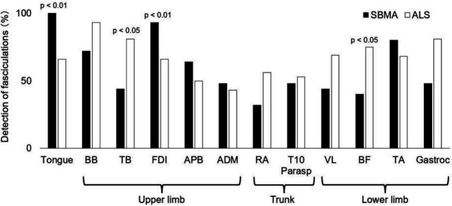
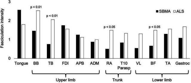
Methods: In 24 SBMA and 16 ALS patients, muscle ultrasonography was systematically performed in the tongue, upper limb muscles (biceps brachii, triceps brachii, first dorsal interosseous (FDI), abductor pollicis brevis and abductor digiti minimi), trunk muscles (Th10 paraspinals and rectus abdominis) and lower limb muscles (vastus lateralis, biceps femoris, tibialis anterior and gastrocnemius). We assessed the presence of fasciculations and the fasciculation intensity (scored from 0 to 3) for each muscle.
Results: All SBMA and ALS patients showed fasciculations at least in two muscles. In SBMA patients, fasciculations were most frequently found in the tongue (100%), FDI (93%) and tibialis anterior (80%), whereas less frequently present in the proximal limb and trunk muscles, irrespective of age, disease duration and CAG repeat numbers. By contrast, in ALS patients, fasciculations were more diffusely distributed including the proximal limb and trunk muscles. When fasciculations were present, the intensity was higher in ALS patients, except for the tongue.
Conclusions: Whereas both diseases exhibit extensive fasciculations, the distribution and intensity are different. SBMA is characterised by prominent involvement in the tongue and distal limb muscles, suggesting different pathophysiology of motor neuronal death in SBMA and ALS.

 求助内容:
求助内容: 应助结果提醒方式:


