Sheng-Yuan Kan, Cinzia G Scarpini, Dawn Ward, Ben Fleming, Heok K Cheow, Ibrahim Jalloh, John A Tadross, James Watkins, Thomas Roberts, Jamie Trotman, Patrick Tarpey, Nicholas Coleman, C Elizabeth Hook, Charlotte Burns, Claire Trayers, Matthew J Murray
{"title":"Mediastinal NUT Carcinoma With Raised Serum Alpha-Fetoprotein Mimicking a Malignant Germ Cell Tumor: Suspicion Raised Due to Negative Serum miR-371a-3p Levels.","authors":"Sheng-Yuan Kan, Cinzia G Scarpini, Dawn Ward, Ben Fleming, Heok K Cheow, Ibrahim Jalloh, John A Tadross, James Watkins, Thomas Roberts, Jamie Trotman, Patrick Tarpey, Nicholas Coleman, C Elizabeth Hook, Charlotte Burns, Claire Trayers, Matthew J Murray","doi":"10.1177/10935266251335391","DOIUrl":null,"url":null,"abstract":"<p><p>NUT carcinoma is challenging to diagnose and may mimic a germ cell tumor (GCT) due to raised serum alpha-fetoprotein (AFP). A 15-year-old patient presented with back pain and cough. Investigation revealed a mediastinal mass and multiple bone metastases. Serum AFP was highly elevated, consistent with a metastatic malignant nonseminomatous GCT. Aggressive chemotherapy was initiated with initial response, unfortunately not sustained. Diagnostic biopsy showed undifferentiated tumor cells with weak GCT immunophenotype but was ultimately non-diagnostic. Serum miR-371a-3p levels, highly sensitive/specific for malignant GCTs, were negative casting diagnostic suspicion. Routine use of agnostic molecular investigations, including whole genome sequencing, identified a chromosome 15:19 translocation, with <i>BRD4::NUTM1</i> gene fusion on RNA sequencing, confirming NUT carcinoma. Subsequent NUTM1 immunohistochemistry was positive. A high index of clinical suspicion is required for non-pathologically/molecularly confirmed diagnoses. Serum miR-371a-3p quantification ruled out malignant GCT and routine agnostic molecular studies identified the correct diagnosis; a low threshold for NUTM1 immunohistochemistry is thus recommended.</p>","PeriodicalId":54634,"journal":{"name":"Pediatric and Developmental Pathology","volume":" ","pages":"338-345"},"PeriodicalIF":1.3000,"publicationDate":"2025-07-01","publicationTypes":"Journal Article","fieldsOfStudy":null,"isOpenAccess":false,"openAccessPdf":"https://www.ncbi.nlm.nih.gov/pmc/articles/PMC12241692/pdf/","citationCount":"0","resultStr":null,"platform":"Semanticscholar","paperid":null,"PeriodicalName":"Pediatric and Developmental Pathology","FirstCategoryId":"3","ListUrlMain":"https://doi.org/10.1177/10935266251335391","RegionNum":4,"RegionCategory":"医学","ArticlePicture":[],"TitleCN":null,"AbstractTextCN":null,"PMCID":null,"EPubDate":"2025/4/25 0:00:00","PubModel":"Epub","JCR":"Q3","JCRName":"PATHOLOGY","Score":null,"Total":0}
引用次数: 0
Abstract
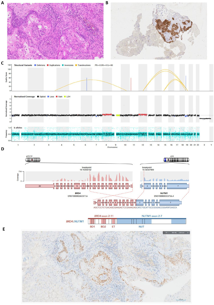
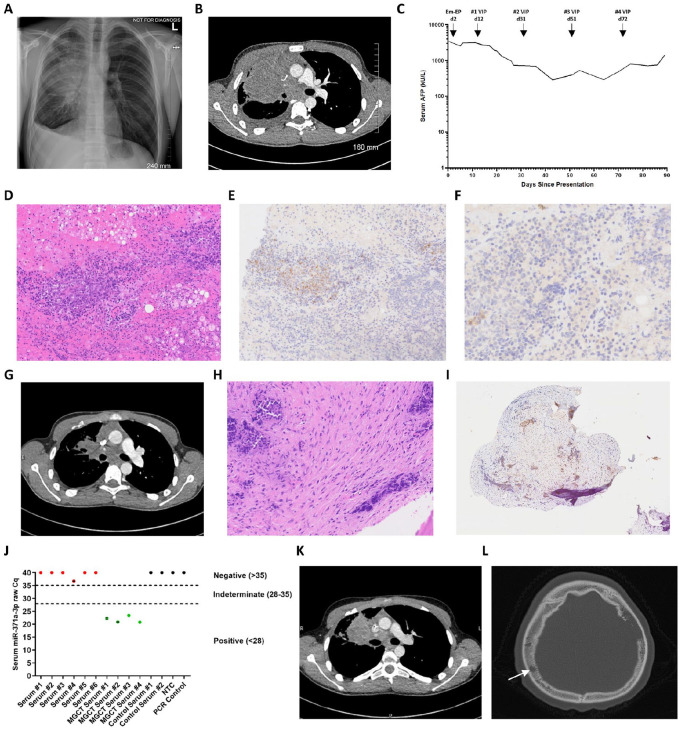
NUT carcinoma is challenging to diagnose and may mimic a germ cell tumor (GCT) due to raised serum alpha-fetoprotein (AFP). A 15-year-old patient presented with back pain and cough. Investigation revealed a mediastinal mass and multiple bone metastases. Serum AFP was highly elevated, consistent with a metastatic malignant nonseminomatous GCT. Aggressive chemotherapy was initiated with initial response, unfortunately not sustained. Diagnostic biopsy showed undifferentiated tumor cells with weak GCT immunophenotype but was ultimately non-diagnostic. Serum miR-371a-3p levels, highly sensitive/specific for malignant GCTs, were negative casting diagnostic suspicion. Routine use of agnostic molecular investigations, including whole genome sequencing, identified a chromosome 15:19 translocation, with BRD4::NUTM1 gene fusion on RNA sequencing, confirming NUT carcinoma. Subsequent NUTM1 immunohistochemistry was positive. A high index of clinical suspicion is required for non-pathologically/molecularly confirmed diagnoses. Serum miR-371a-3p quantification ruled out malignant GCT and routine agnostic molecular studies identified the correct diagnosis; a low threshold for NUTM1 immunohistochemistry is thus recommended.
期刊介绍:
The Journal covers the spectrum of disorders of early development (including embryology, placentology, and teratology), gestational and perinatal diseases, and all diseases of childhood. Studies may be in any field of experimental, anatomic, or clinical pathology, including molecular pathology. Case reports are published only if they provide new insights into disease mechanisms or new information.

 求助内容:
求助内容: 应助结果提醒方式:


