М M Abdurakhmanova, A A Leonteva, N S Vasilieva, E V Kuligina, A A Nushtaeva
{"title":"3D cell culture models: how to obtain and characterize the main models.","authors":"М M Abdurakhmanova, A A Leonteva, N S Vasilieva, E V Kuligina, A A Nushtaeva","doi":"10.18699/vjgb-25-21","DOIUrl":null,"url":null,"abstract":"<p><p>For many years, the gold standard in the study of malignant tumors has been the in vitro culture of tumor cells, in vivo xenografts or genetically modified animal models. Meanwhile, three-dimensional cell models (3D cultures) have been added to the arsenal of modern biomedical research. 3D cultures reproduce tissue-specific features of tissue topology. This makes them relevant tissue models in terms of cell differentiation, metabolism and the development of drug resistance. Such models are already being used by many research groups for both basic and translational research, and may substantially reduce the number of animal studies, for example in the field of oncological research. In the current literature, 3D cultures are classified according to the technique of their formation (with or without a scaffold), cultivation conditions (static or dynamic), as well as their cellular organization and function. In terms of cellular organization, 3D cultures are divided into \"spheroid models\", \"organoids\", \"organs-on-a-chip\" and \"microtissues\". Each of these models has its own unique features, which should be taken into account when using a particular model in an experiment. The simplest 3D cultures are spheroid models which are floating spherical cell aggregates. An organoid is a more complex 3D model, in which a self-organizing 3D structure is formed from stem cells (SCs) capable of self-renewal and differentiation within the model. Organ-on-a-chip models are chips of microfluidic systems that simulate dynamic physical and biological processes found in organs and tissues in vitro. By combining different cell types into a single structure, spheroids and organoids can act as a basis for the formation of a microtissue - a hybrid 3D model imitating a specific tissue phenotype and containing tissue-specific extracellular matrix (ECM) components. This review presents a brief history of 3D cell culture. It describes the main characteristics and perspectives of the use of \"spheroid models\", \"organoids\", \"organ-on-a-chip\" models and \"microtissues\" in immune oncology research of solid tumors.</p>","PeriodicalId":44339,"journal":{"name":"Vavilovskii Zhurnal Genetiki i Selektsii","volume":"29 2","pages":"175-188"},"PeriodicalIF":1.0000,"publicationDate":"2025-04-01","publicationTypes":"Journal Article","fieldsOfStudy":null,"isOpenAccess":false,"openAccessPdf":"https://www.ncbi.nlm.nih.gov/pmc/articles/PMC12011624/pdf/","citationCount":"0","resultStr":null,"platform":"Semanticscholar","paperid":null,"PeriodicalName":"Vavilovskii Zhurnal Genetiki i Selektsii","FirstCategoryId":"1085","ListUrlMain":"https://doi.org/10.18699/vjgb-25-21","RegionNum":0,"RegionCategory":null,"ArticlePicture":[],"TitleCN":null,"AbstractTextCN":null,"PMCID":null,"EPubDate":"","PubModel":"","JCR":"Q3","JCRName":"AGRICULTURE, MULTIDISCIPLINARY","Score":null,"Total":0}
引用次数: 0
Abstract
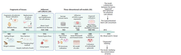
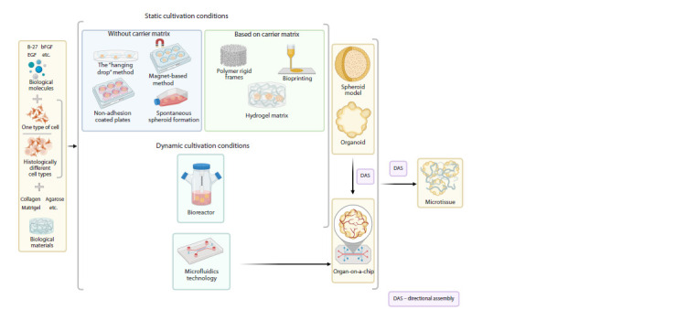
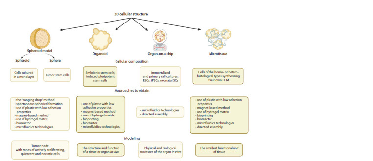
For many years, the gold standard in the study of malignant tumors has been the in vitro culture of tumor cells, in vivo xenografts or genetically modified animal models. Meanwhile, three-dimensional cell models (3D cultures) have been added to the arsenal of modern biomedical research. 3D cultures reproduce tissue-specific features of tissue topology. This makes them relevant tissue models in terms of cell differentiation, metabolism and the development of drug resistance. Such models are already being used by many research groups for both basic and translational research, and may substantially reduce the number of animal studies, for example in the field of oncological research. In the current literature, 3D cultures are classified according to the technique of their formation (with or without a scaffold), cultivation conditions (static or dynamic), as well as their cellular organization and function. In terms of cellular organization, 3D cultures are divided into "spheroid models", "organoids", "organs-on-a-chip" and "microtissues". Each of these models has its own unique features, which should be taken into account when using a particular model in an experiment. The simplest 3D cultures are spheroid models which are floating spherical cell aggregates. An organoid is a more complex 3D model, in which a self-organizing 3D structure is formed from stem cells (SCs) capable of self-renewal and differentiation within the model. Organ-on-a-chip models are chips of microfluidic systems that simulate dynamic physical and biological processes found in organs and tissues in vitro. By combining different cell types into a single structure, spheroids and organoids can act as a basis for the formation of a microtissue - a hybrid 3D model imitating a specific tissue phenotype and containing tissue-specific extracellular matrix (ECM) components. This review presents a brief history of 3D cell culture. It describes the main characteristics and perspectives of the use of "spheroid models", "organoids", "organ-on-a-chip" models and "microtissues" in immune oncology research of solid tumors.
期刊介绍:
The "Vavilov Journal of genetics and breeding" publishes original research and review articles in all key areas of modern plant, animal and human genetics, genomics, bioinformatics and biotechnology. One of the main objectives of the journal is integration of theoretical and applied research in the field of genetics. Special attention is paid to the most topical areas in modern genetics dealing with global concerns such as food security and human health.

 求助内容:
求助内容: 应助结果提醒方式:


