{"title":"A rare cause of knee pain: Multiple intraosseous lipomas mimicking bone metastasis.","authors":"Hatice Betigul Meral, Mehmet Agirman","doi":"10.52312/jdrs.2025.2017","DOIUrl":null,"url":null,"abstract":"<p><p>Intraosseous lipomas (IOLs) are rare benign bone tumors which can closely mimic malignancy on imaging, leading to diagnostic challenges. A 34-year-old male patient was admitted with persistent knee pain which was initially suspected to be metastatic bone disease. The patient reported activity-related pain relieved by rest. Physical examination revealed mild tenderness without swelling or restricted motion. Laboratory findings, including serum calcium and inflammatory markers, were normal. Magnetic resonance imaging (MRI) showed hyperintense lesions in the tibial and femoral metaphyses with surrounding bone marrow edema, raising suspicion of malignancy. Bone scintigraphy revealed increased radionuclide uptake in the tibial tuberosity, iliac crest, and the ninth rib, further mimicking metastatic disease. A biopsy of the tibial lesion confirmed mature adipocytes without necrosis or atypia, diagnosing IOLs. The patient was managed conservatively with non-steroidal anti-inflammatory drugs and physiotherapy. At three months, symptoms resolved completely, with no recurrence or new complaints. Follow-up MRI confirmed lesion stability. In conclusion, this case highlights the diagnostic complexity of IOLs and emphasizes the need for multimodal imaging and histopathological confirmation to differentiate benign lesions from malignancies. Including IOLs in the differential diagnosis of unexplained bone pain can prevent unnecessary invasive procedures and mistreatment.</p>","PeriodicalId":73560,"journal":{"name":"Joint diseases and related surgery","volume":"36 2","pages":"467-471"},"PeriodicalIF":1.9000,"publicationDate":"2025-04-05","publicationTypes":"Journal Article","fieldsOfStudy":null,"isOpenAccess":false,"openAccessPdf":"https://www.ncbi.nlm.nih.gov/pmc/articles/PMC12086487/pdf/","citationCount":"0","resultStr":null,"platform":"Semanticscholar","paperid":null,"PeriodicalName":"Joint diseases and related surgery","FirstCategoryId":"1085","ListUrlMain":"https://doi.org/10.52312/jdrs.2025.2017","RegionNum":0,"RegionCategory":null,"ArticlePicture":[],"TitleCN":null,"AbstractTextCN":null,"PMCID":null,"EPubDate":"","PubModel":"","JCR":"Q2","JCRName":"ORTHOPEDICS","Score":null,"Total":0}
引用次数: 0
Abstract
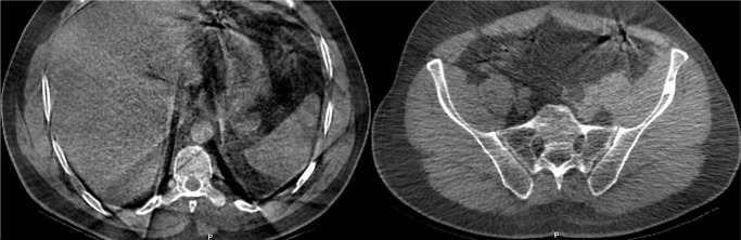
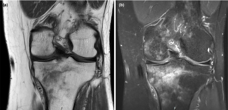
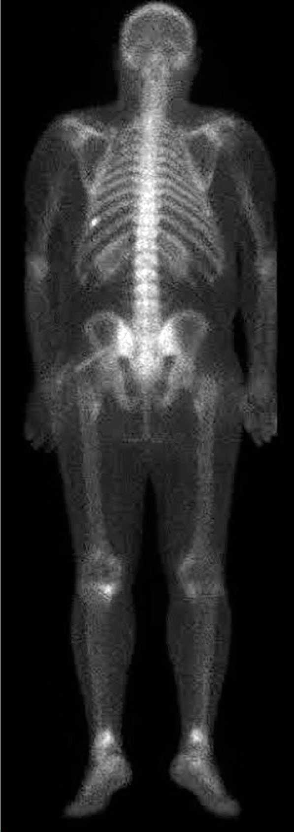
Intraosseous lipomas (IOLs) are rare benign bone tumors which can closely mimic malignancy on imaging, leading to diagnostic challenges. A 34-year-old male patient was admitted with persistent knee pain which was initially suspected to be metastatic bone disease. The patient reported activity-related pain relieved by rest. Physical examination revealed mild tenderness without swelling or restricted motion. Laboratory findings, including serum calcium and inflammatory markers, were normal. Magnetic resonance imaging (MRI) showed hyperintense lesions in the tibial and femoral metaphyses with surrounding bone marrow edema, raising suspicion of malignancy. Bone scintigraphy revealed increased radionuclide uptake in the tibial tuberosity, iliac crest, and the ninth rib, further mimicking metastatic disease. A biopsy of the tibial lesion confirmed mature adipocytes without necrosis or atypia, diagnosing IOLs. The patient was managed conservatively with non-steroidal anti-inflammatory drugs and physiotherapy. At three months, symptoms resolved completely, with no recurrence or new complaints. Follow-up MRI confirmed lesion stability. In conclusion, this case highlights the diagnostic complexity of IOLs and emphasizes the need for multimodal imaging and histopathological confirmation to differentiate benign lesions from malignancies. Including IOLs in the differential diagnosis of unexplained bone pain can prevent unnecessary invasive procedures and mistreatment.

 求助内容:
求助内容: 应助结果提醒方式:


