Abscisic Acid Rescues Behavior in Adult Female Mice in Attention Deficit Disorder with Hyperactivity Model of Dopamine Depletion by Regulating Microglia and Increasing Vesicular GABA Transporter Expression.
Maria Meseguer-Beltrán, Sandra Sánchez-Sarasúa, Nóra Kerekes, Marc Landry, Matías Real-López, Ana María Sánchez-Pérez
{"title":"Abscisic Acid Rescues Behavior in Adult Female Mice in Attention Deficit Disorder with Hyperactivity Model of Dopamine Depletion by Regulating Microglia and Increasing Vesicular GABA Transporter Expression.","authors":"Maria Meseguer-Beltrán, Sandra Sánchez-Sarasúa, Nóra Kerekes, Marc Landry, Matías Real-López, Ana María Sánchez-Pérez","doi":"10.1007/s11481-025-10186-6","DOIUrl":null,"url":null,"abstract":"<p><p>Attention-deficit/hyperactivity disorder (ADHD) is a neurodevelopmental syndrome typically diagnosed in childhood that may persist into adulthood. Its etiology encompasses both genetic and environmental factors, with genetic studies indicating catecholamine dysfunction and epidemiological evidence emphasizing neuroinflammation as a potential trigger. To investigate the roles of inflammation and development processes in ADHD, we conducted a longitudinal behavioral study using female Swiss mice with a dopamine deficit model. We explored the impact of neonatal dopaminergic lesions, treatment with abscisic acid (ABA)-an anti-inflammatory hormone-and developmental changes by comparing behavioral patterns in juvenile and adult mice. Postmortem analyses assessed neuroinflammation through microglial morphology, NLRP3, cytokine expression, and the excitatory/inhibitory (E/I) ratio in specific brain regions. Neonatal dopaminergic lesions induced hyperactivity and hypersensitivity in juvenile mice that persisted into adulthood. In adults, increased social interaction and memory impairment were observed in lesioned mice. Brain development mitigated impulsivity, while ABA treatment reduced locomotor activity, downregulated pain sensitivity, and influenced social interaction, although it did not completely resolve cognitive deficits in lesioned adult mice. In brain regions such as the anterior cingulate cortex (ACC), posterior insular cortex (pIC), and hippocampus, lesions significantly altered microglial morphology. In the ACC, lesions increased IL-1β and TNFα levels, decreased Arg1 mRNA levels, and disrupted the E/I balance. Importantly, ABA treatment restored microglial morphology, normalized IL-1β and Arg1 expression and upregulated vGAT levels. This study demonstrates that dopamine deficits lead to microglia alterations and E/I imbalance, contributing to ADHD symptoms. While some symptoms improve with brain development, targeting microglial health in specific brain regions emerges as a promising therapeutic approach for managing ADHD.</p>","PeriodicalId":73858,"journal":{"name":"Journal of neuroimmune pharmacology : the official journal of the Society on NeuroImmune Pharmacology","volume":"20 1","pages":"39"},"PeriodicalIF":3.5000,"publicationDate":"2025-04-16","publicationTypes":"Journal Article","fieldsOfStudy":null,"isOpenAccess":false,"openAccessPdf":"https://www.ncbi.nlm.nih.gov/pmc/articles/PMC12000189/pdf/","citationCount":"0","resultStr":null,"platform":"Semanticscholar","paperid":null,"PeriodicalName":"Journal of neuroimmune pharmacology : the official journal of the Society on NeuroImmune Pharmacology","FirstCategoryId":"1085","ListUrlMain":"https://doi.org/10.1007/s11481-025-10186-6","RegionNum":0,"RegionCategory":null,"ArticlePicture":[],"TitleCN":null,"AbstractTextCN":null,"PMCID":null,"EPubDate":"","PubModel":"","JCR":"","JCRName":"","Score":null,"Total":0}
引用次数: 0
Abstract
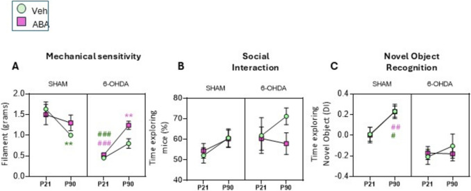
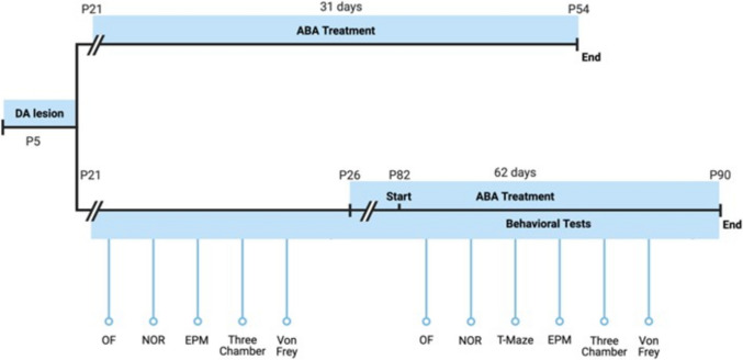
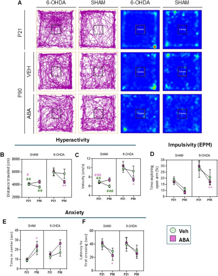
Attention-deficit/hyperactivity disorder (ADHD) is a neurodevelopmental syndrome typically diagnosed in childhood that may persist into adulthood. Its etiology encompasses both genetic and environmental factors, with genetic studies indicating catecholamine dysfunction and epidemiological evidence emphasizing neuroinflammation as a potential trigger. To investigate the roles of inflammation and development processes in ADHD, we conducted a longitudinal behavioral study using female Swiss mice with a dopamine deficit model. We explored the impact of neonatal dopaminergic lesions, treatment with abscisic acid (ABA)-an anti-inflammatory hormone-and developmental changes by comparing behavioral patterns in juvenile and adult mice. Postmortem analyses assessed neuroinflammation through microglial morphology, NLRP3, cytokine expression, and the excitatory/inhibitory (E/I) ratio in specific brain regions. Neonatal dopaminergic lesions induced hyperactivity and hypersensitivity in juvenile mice that persisted into adulthood. In adults, increased social interaction and memory impairment were observed in lesioned mice. Brain development mitigated impulsivity, while ABA treatment reduced locomotor activity, downregulated pain sensitivity, and influenced social interaction, although it did not completely resolve cognitive deficits in lesioned adult mice. In brain regions such as the anterior cingulate cortex (ACC), posterior insular cortex (pIC), and hippocampus, lesions significantly altered microglial morphology. In the ACC, lesions increased IL-1β and TNFα levels, decreased Arg1 mRNA levels, and disrupted the E/I balance. Importantly, ABA treatment restored microglial morphology, normalized IL-1β and Arg1 expression and upregulated vGAT levels. This study demonstrates that dopamine deficits lead to microglia alterations and E/I imbalance, contributing to ADHD symptoms. While some symptoms improve with brain development, targeting microglial health in specific brain regions emerges as a promising therapeutic approach for managing ADHD.

 求助内容:
求助内容: 应助结果提醒方式:


