Gehad Ahmad Saleh, Omar Hamdy, Dina Ragab, Bassante Farouk, Mennatalla Mahmoud Allam, Rawan Abo Asy, Fatmaelzahraa A Denewar, Mohamed Ezat
{"title":"Fibrothecoma of the Ovary; Clinical and Imaging Characteristics.","authors":"Gehad Ahmad Saleh, Omar Hamdy, Dina Ragab, Bassante Farouk, Mennatalla Mahmoud Allam, Rawan Abo Asy, Fatmaelzahraa A Denewar, Mohamed Ezat","doi":"10.1089/whr.2024.0153","DOIUrl":null,"url":null,"abstract":"<p><strong>Introduction: </strong>Ovarian fibrothecoma is a rare benign sex cord-stromal ovarian tumor sorted under the thecoma-fibroma group. We present an analysis of clinical and laboratory findings and the radiological characteristic features of pathologically proven fibrothecomas in variable imaging modalities.</p><p><strong>Methods: </strong>A retrospective analysis was done for 88 patients with 90 pathologically proven ovarian fibrothecoma between January 2011 and December 2023 from our center's prospectively maintained database. All the patients underwent preoperative ultrasonography, computed tomography (CT), and magnetic resonance imaging (MRI) scans, clinical examinations, basic laboratory tests, and tumor markers.</p><p><strong>Results: </strong>The results of Spearman's correlation revealed a statistically significant positive correlation between the largest tumor diameter and serum level. CA 125, the degree of ascites, and diffusion weighted imaging (DWI) signal intensity while the results of point biserial correlation revealed a statistically significant correlation of the largest tumor diameter with the presence of ascites, cystic changes, abdominal enlargement, surgery type, and border type. There were also statistically significantly higher hypoechoic lesions in the smaller tumor group (<i>p</i> = 0.001) but not for isoechoic (<i>p</i> = 0.099) and mixed (<i>p</i> = 0.052). Regarding the MRI, there was a statistically significantly larger tumor diameter in T2 mixed-hyperintense versus hypointense (<i>p</i> = 0.007) and intermediate (<i>p</i> = 0.010) signal intensities.</p><p><strong>Conclusion: </strong>Fibrothecoma showed a statistically significant positive correlation between the largest tumor diameter with serum level CA 125 and the amount of ascites. On imaging, it shows mild enhancement in both CT and MRI, with a statistically significant positive correlation of the largest tumor diameter with T2 and DWI signal intensity.</p>","PeriodicalId":75329,"journal":{"name":"Women's health reports (New Rochelle, N.Y.)","volume":"6 1","pages":"315-324"},"PeriodicalIF":1.8000,"publicationDate":"2025-03-25","publicationTypes":"Journal Article","fieldsOfStudy":null,"isOpenAccess":false,"openAccessPdf":"https://www.ncbi.nlm.nih.gov/pmc/articles/PMC12040543/pdf/","citationCount":"0","resultStr":null,"platform":"Semanticscholar","paperid":null,"PeriodicalName":"Women's health reports (New Rochelle, N.Y.)","FirstCategoryId":"1085","ListUrlMain":"https://doi.org/10.1089/whr.2024.0153","RegionNum":0,"RegionCategory":null,"ArticlePicture":[],"TitleCN":null,"AbstractTextCN":null,"PMCID":null,"EPubDate":"2025/1/1 0:00:00","PubModel":"eCollection","JCR":"Q3","JCRName":"OBSTETRICS & GYNECOLOGY","Score":null,"Total":0}
引用次数: 0
Abstract
Introduction: Ovarian fibrothecoma is a rare benign sex cord-stromal ovarian tumor sorted under the thecoma-fibroma group. We present an analysis of clinical and laboratory findings and the radiological characteristic features of pathologically proven fibrothecomas in variable imaging modalities.
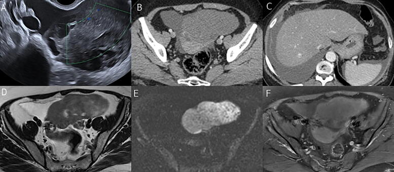
Methods: A retrospective analysis was done for 88 patients with 90 pathologically proven ovarian fibrothecoma between January 2011 and December 2023 from our center's prospectively maintained database. All the patients underwent preoperative ultrasonography, computed tomography (CT), and magnetic resonance imaging (MRI) scans, clinical examinations, basic laboratory tests, and tumor markers.
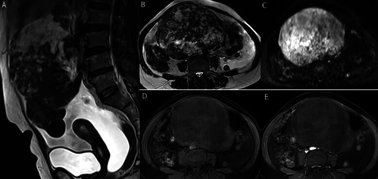
Results: The results of Spearman's correlation revealed a statistically significant positive correlation between the largest tumor diameter and serum level. CA 125, the degree of ascites, and diffusion weighted imaging (DWI) signal intensity while the results of point biserial correlation revealed a statistically significant correlation of the largest tumor diameter with the presence of ascites, cystic changes, abdominal enlargement, surgery type, and border type. There were also statistically significantly higher hypoechoic lesions in the smaller tumor group (p = 0.001) but not for isoechoic (p = 0.099) and mixed (p = 0.052). Regarding the MRI, there was a statistically significantly larger tumor diameter in T2 mixed-hyperintense versus hypointense (p = 0.007) and intermediate (p = 0.010) signal intensities.
Conclusion: Fibrothecoma showed a statistically significant positive correlation between the largest tumor diameter with serum level CA 125 and the amount of ascites. On imaging, it shows mild enhancement in both CT and MRI, with a statistically significant positive correlation of the largest tumor diameter with T2 and DWI signal intensity.

 求助内容:
求助内容: 应助结果提醒方式:


