The Prognostic Value of Body Composition Analysis on Non-Enhanced CT for Risk Stratification in Gastrointestinal Stromal Tumors: A Retrospective Study.
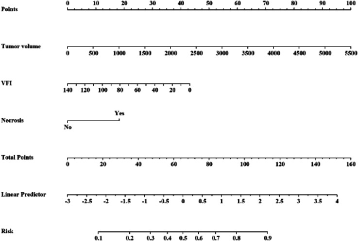
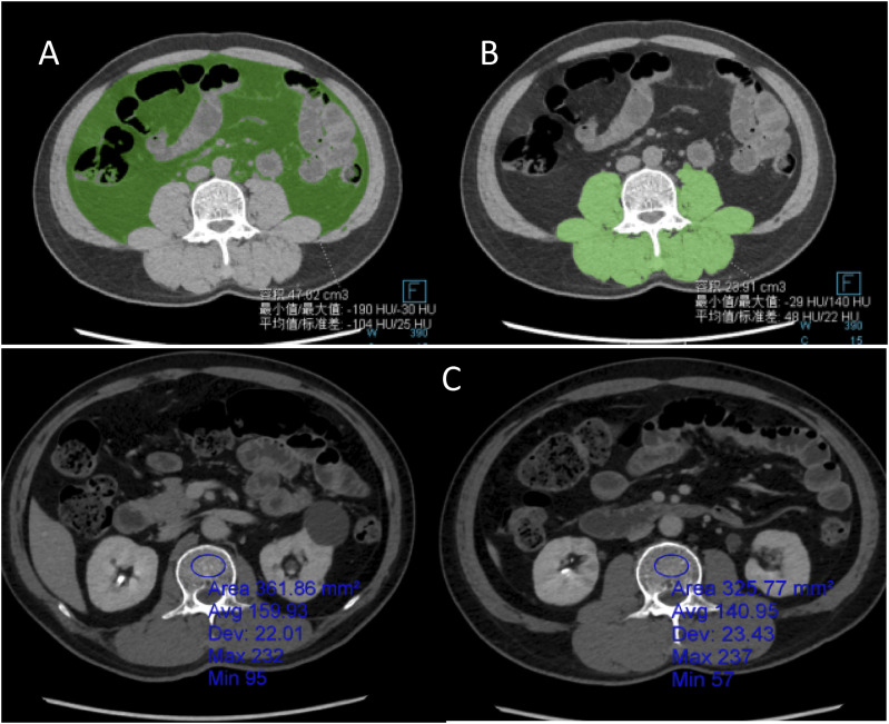
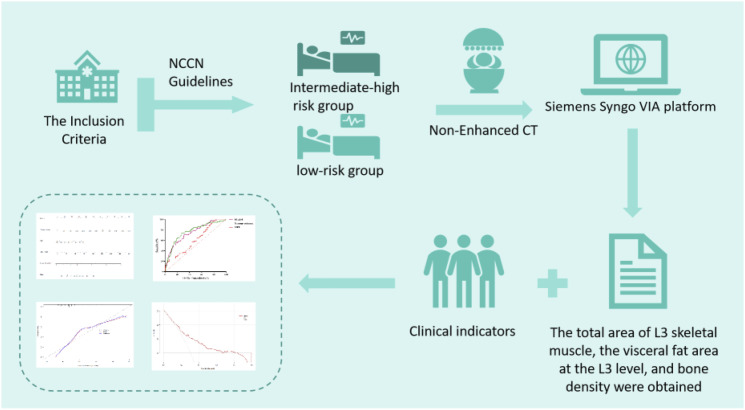
{"title":"The Prognostic Value of Body Composition Analysis on Non-Enhanced CT for Risk Stratification in Gastrointestinal Stromal Tumors: A Retrospective Study.","authors":"Wei Chen, Long-Yu Duan, Xiao-Juan Peng, Kun-Ming Yi, Lian-Qin Kuang","doi":"10.1177/10732748251342068","DOIUrl":null,"url":null,"abstract":"<p><p>IntroductionContrast-enhanced computed tomography (CT) is the primary imaging modality for accurate risk stratification in gastrointestinal stromal tumors (GISTs). However, contrast-enhanced CT may not always be accessible or suitable for all patients undergoing risk assessment of GISTs. Therefore, this study explored the use of non-enhanced CT imaging for assessing body composition in patients with GISTs to preoperatively predict risk stratification.MethodsWe retrospectively analyzed 233 patients with GISTs who met the inclusion criteria. Pretreatment complete abdominal CT images from these patients were processed and analyzed using the Siemens Syngo imaging system. The data were subsequently organized and analyzed using the SPSS software (version 26.0).ResultsThrough two independent samples t-tests, Mann-Whitney U tests, and chi-square tests (including corrected chi-square tests and Fisher's exact tests), the intermediate-high risk group exhibited a lower visceral fat index (VFI) and higher tumor volumes and proportions of necrosis (<i>P</i> < .05), compared to the low-risk group (<i>P</i> < .05). No statistically significant differences were observed in the other indicators. Our research demonstrates that tumor volume is positively correlated with the National Institutes of Health (NIH) classification and exhibits the highest specificity among the four models (specificity = 0.735). However, its sensitivity is lower than that of the combined model (sensitivity = 0.803) and the VFI model (sensitivity = 0.972).ConclusionBased on the vascular abundance index, tumor volume, and necrosis status observed in the CT plain scan images of patients with GIST, a comprehensive predictive model was developed. This model can accurately predict the NIH grade of stromal tumors, thereby providing a robust basis for formulating effective treatment strategies and improving the prognosis of patients with GISTs who cannot undergo contrast-enhanced CT.</p>","PeriodicalId":49093,"journal":{"name":"Cancer Control","volume":"32 ","pages":"10732748251342068"},"PeriodicalIF":2.6000,"publicationDate":"2025-01-01","publicationTypes":"Journal Article","fieldsOfStudy":null,"isOpenAccess":false,"openAccessPdf":"https://www.ncbi.nlm.nih.gov/pmc/articles/PMC12066848/pdf/","citationCount":"0","resultStr":null,"platform":"Semanticscholar","paperid":null,"PeriodicalName":"Cancer Control","FirstCategoryId":"3","ListUrlMain":"https://doi.org/10.1177/10732748251342068","RegionNum":4,"RegionCategory":"医学","ArticlePicture":[],"TitleCN":null,"AbstractTextCN":null,"PMCID":null,"EPubDate":"2025/5/11 0:00:00","PubModel":"Epub","JCR":"Q3","JCRName":"ONCOLOGY","Score":null,"Total":0}
引用次数: 0
Abstract
IntroductionContrast-enhanced computed tomography (CT) is the primary imaging modality for accurate risk stratification in gastrointestinal stromal tumors (GISTs). However, contrast-enhanced CT may not always be accessible or suitable for all patients undergoing risk assessment of GISTs. Therefore, this study explored the use of non-enhanced CT imaging for assessing body composition in patients with GISTs to preoperatively predict risk stratification.MethodsWe retrospectively analyzed 233 patients with GISTs who met the inclusion criteria. Pretreatment complete abdominal CT images from these patients were processed and analyzed using the Siemens Syngo imaging system. The data were subsequently organized and analyzed using the SPSS software (version 26.0).ResultsThrough two independent samples t-tests, Mann-Whitney U tests, and chi-square tests (including corrected chi-square tests and Fisher's exact tests), the intermediate-high risk group exhibited a lower visceral fat index (VFI) and higher tumor volumes and proportions of necrosis (P < .05), compared to the low-risk group (P < .05). No statistically significant differences were observed in the other indicators. Our research demonstrates that tumor volume is positively correlated with the National Institutes of Health (NIH) classification and exhibits the highest specificity among the four models (specificity = 0.735). However, its sensitivity is lower than that of the combined model (sensitivity = 0.803) and the VFI model (sensitivity = 0.972).ConclusionBased on the vascular abundance index, tumor volume, and necrosis status observed in the CT plain scan images of patients with GIST, a comprehensive predictive model was developed. This model can accurately predict the NIH grade of stromal tumors, thereby providing a robust basis for formulating effective treatment strategies and improving the prognosis of patients with GISTs who cannot undergo contrast-enhanced CT.
期刊介绍:
Cancer Control is a JCR-ranked, peer-reviewed open access journal whose mission is to advance the prevention, detection, diagnosis, treatment, and palliative care of cancer by enabling researchers, doctors, policymakers, and other healthcare professionals to freely share research along the cancer control continuum. Our vision is a world where gold-standard cancer care is the norm, not the exception.
 求助内容:
求助内容: 应助结果提醒方式:


