Bernardo Lopes Crisostomo, Julia Pozzetti Daou, Jairo Greco Garcia, Marcelo DE Toledo Petrilli, Dan Carai Maia Viola, Reynaldo Jesus Garcia
{"title":"EPIDEMOLOGICAL, RADIOGRAPHIC AND PROGNOSTIC EVALUATION OF CHONDROBLASTOMA.","authors":"Bernardo Lopes Crisostomo, Julia Pozzetti Daou, Jairo Greco Garcia, Marcelo DE Toledo Petrilli, Dan Carai Maia Viola, Reynaldo Jesus Garcia","doi":"10.1590/1413-785220243201e283605","DOIUrl":null,"url":null,"abstract":"<p><strong>Objective: </strong>To describe the clinical and imaging characteristics of chondroblastoma and identify possible factors related to joint complications.</p><p><strong>Method: </strong>This retrospective cohort study was carried out with data from the medical records of 23 patients diagnosed with chondroblastoma, subjecting them to statistical analyses.</p><p><strong>Result: </strong>In total, 19 patients were included, 12 (63.2%) of which were mean with a mean age of 13.6±3.5 year. The relation with the local dimension equaled 57.9%, higher in the apophysis of the greater trochanter: 95.2% (p<0.001). Based on imaging, 15.8% patients had an open physis; 55.6%, no damaged physeal line; 42.1%, cortical rupture; 21.1%, secondary aneurysmal bone cyst; 26.7%, violated cartilage; and all cases, medullary edema. 15.8% of cases showed local recurrence and no metastasis. Moreover, 46.7% of patients had relevant secondary osteoarthritis related to the aggressiveness of the tumor according to the Enneking classification (p= 0.041).</p><p><strong>Conclusion: </strong>The clinical outcome of chondroblastoma show no relation to age, sex, location, physeal status, or presence of calcifications or secondary aneurysmal bone cyst. Progression to secondary osteoarthritis configured the most frequent non-oncological complication and showed a direct relation with the severity of the chondroblastoma. <b><i>Level of Evidence IV, Case Series.</i></b></p>","PeriodicalId":55563,"journal":{"name":"Acta Ortopedica Brasileira","volume":"33 spe1","pages":"e283605"},"PeriodicalIF":0.6000,"publicationDate":"2025-04-07","publicationTypes":"Journal Article","fieldsOfStudy":null,"isOpenAccess":false,"openAccessPdf":"https://www.ncbi.nlm.nih.gov/pmc/articles/PMC11978305/pdf/","citationCount":"0","resultStr":null,"platform":"Semanticscholar","paperid":null,"PeriodicalName":"Acta Ortopedica Brasileira","FirstCategoryId":"3","ListUrlMain":"https://doi.org/10.1590/1413-785220243201e283605","RegionNum":4,"RegionCategory":"医学","ArticlePicture":[],"TitleCN":null,"AbstractTextCN":null,"PMCID":null,"EPubDate":"2025/1/1 0:00:00","PubModel":"eCollection","JCR":"Q4","JCRName":"ORTHOPEDICS","Score":null,"Total":0}
引用次数: 0
Abstract
Objective: To describe the clinical and imaging characteristics of chondroblastoma and identify possible factors related to joint complications.
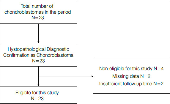
Method: This retrospective cohort study was carried out with data from the medical records of 23 patients diagnosed with chondroblastoma, subjecting them to statistical analyses.
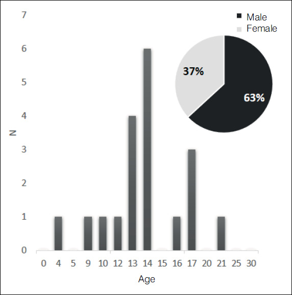
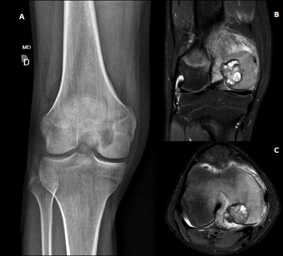
Result: In total, 19 patients were included, 12 (63.2%) of which were mean with a mean age of 13.6±3.5 year. The relation with the local dimension equaled 57.9%, higher in the apophysis of the greater trochanter: 95.2% (p<0.001). Based on imaging, 15.8% patients had an open physis; 55.6%, no damaged physeal line; 42.1%, cortical rupture; 21.1%, secondary aneurysmal bone cyst; 26.7%, violated cartilage; and all cases, medullary edema. 15.8% of cases showed local recurrence and no metastasis. Moreover, 46.7% of patients had relevant secondary osteoarthritis related to the aggressiveness of the tumor according to the Enneking classification (p= 0.041).
Conclusion: The clinical outcome of chondroblastoma show no relation to age, sex, location, physeal status, or presence of calcifications or secondary aneurysmal bone cyst. Progression to secondary osteoarthritis configured the most frequent non-oncological complication and showed a direct relation with the severity of the chondroblastoma. Level of Evidence IV, Case Series.
期刊介绍:
A Revista Acta Ortopédica Brasileira, órgão oficial do Departamento de Ortopedia e Traumatologia da Faculdade de Medicina da Universidade de São Paulo (DOT/FMUSP), é publicada bimestralmente em seis edições ao ano (jan/fev, mar/abr, maio/jun, jul/ago, set/out e nov/dez) com versão em inglês disponível nos principais indexadores nacionais e internacionais e instituições de ensino do Brasil. Sendo hoje reconhecidamente uma importante contribuição para os especialistas da área com sua seriedade e árduo trabalho para as indexações já conquistadas.
 求助内容:
求助内容: 应助结果提醒方式:


