Ola Sobhy A Elmeseiny, Simon Winther, Hanne Skou Jørgensen, My Svensson, Morten Bøttcher, Per Ivarsen, Gratien Andersen, Henrik Birn, Marie Bodilsen Nielsen
{"title":"Assessment of aortic and iliac artery calcification using CT-angiography in kidney transplant candidates.","authors":"Ola Sobhy A Elmeseiny, Simon Winther, Hanne Skou Jørgensen, My Svensson, Morten Bøttcher, Per Ivarsen, Gratien Andersen, Henrik Birn, Marie Bodilsen Nielsen","doi":"10.1186/s42155-025-00542-1","DOIUrl":null,"url":null,"abstract":"<p><strong>Purpose: </strong>Assessment of vascular calcification provides the opportunity for risk stratification in kidney transplant candidates (KTCs), as vascular calcification constitutes an independent risk factor for cardiovascular events. The aim of the present study is to explore the feasibility of contrast enhanced computed tomography (CT)-angiography to quantitate vascular calcification, to avoid the extra radiation of an additional non-contrast CT scan.</p><p><strong>Methods and materials: </strong>43 KTCs who underwent concomitant non-contrast CT scans and CT-angiographies of the infrarenal aorta and iliac arteries were included. Vascular calcification was quantified using the Agatston method on non-contrast CT and applying individual Hounsfield Unit thresholds on CT-angiographies based on the radio density of the aortic lumen. The calcium scores and volumes from non-contrast CT scans and CT-angiographies were compared using linear regression and Bland-Altman plots.</p><p><strong>Results: </strong>Non-contrast CT revealed vascular calcification in the infrarenal aorta in 92% of KTCs and in the iliac arteries in 90% of KTCs. The calcium scores estimated from CT-angiography correlated linearly with the calcium scores based on non-contrast CT scans (infrarenal aorta: R<sup>2</sup> = 0.71, p < 0.0001; iliac arteries: R<sup>2</sup> = 0.71, p < 0.0001); however, the calcium scores were higher, and volumes were lower compared to the non-contrast CT scans. The median differences in calcium scores were 1517 [48 - 6138] for the infrarenal aorta, and 2361 [59 - 8644] for the iliac arteries.</p><p><strong>Conclusion: </strong>Vascular calcification is present in the majority of KTCs. Calcification of the infrarenal aorta and iliac arteries may be assessed using CT-angiography, though higher calcium scores and lower volumes are found compared to the non-contrast CT scan.</p>","PeriodicalId":52351,"journal":{"name":"CVIR Endovascular","volume":"8 1","pages":"39"},"PeriodicalIF":1.5000,"publicationDate":"2025-05-06","publicationTypes":"Journal Article","fieldsOfStudy":null,"isOpenAccess":false,"openAccessPdf":"https://www.ncbi.nlm.nih.gov/pmc/articles/PMC12055728/pdf/","citationCount":"0","resultStr":null,"platform":"Semanticscholar","paperid":null,"PeriodicalName":"CVIR Endovascular","FirstCategoryId":"1085","ListUrlMain":"https://doi.org/10.1186/s42155-025-00542-1","RegionNum":0,"RegionCategory":null,"ArticlePicture":[],"TitleCN":null,"AbstractTextCN":null,"PMCID":null,"EPubDate":"","PubModel":"","JCR":"Q3","JCRName":"CARDIAC & CARDIOVASCULAR SYSTEMS","Score":null,"Total":0}
引用次数: 0
Abstract
Purpose: Assessment of vascular calcification provides the opportunity for risk stratification in kidney transplant candidates (KTCs), as vascular calcification constitutes an independent risk factor for cardiovascular events. The aim of the present study is to explore the feasibility of contrast enhanced computed tomography (CT)-angiography to quantitate vascular calcification, to avoid the extra radiation of an additional non-contrast CT scan.
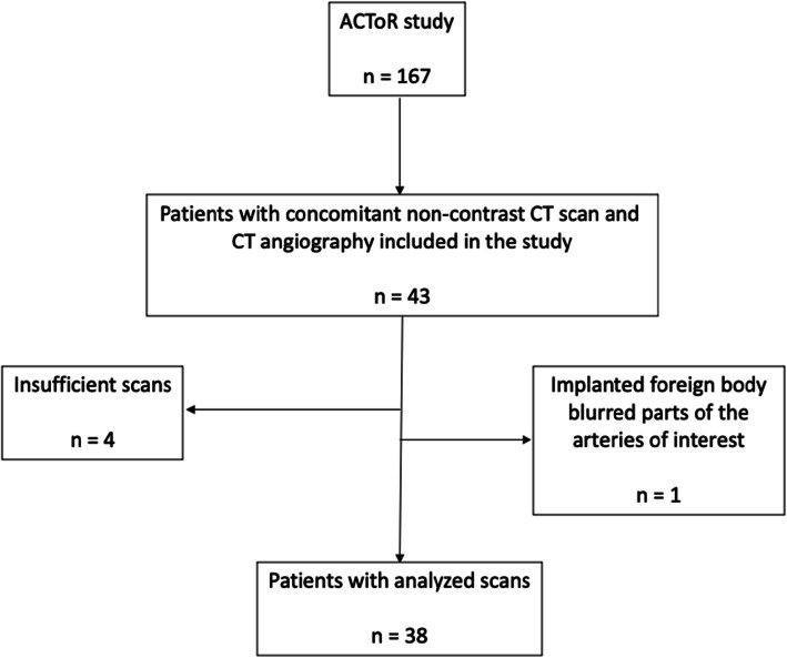
Methods and materials: 43 KTCs who underwent concomitant non-contrast CT scans and CT-angiographies of the infrarenal aorta and iliac arteries were included. Vascular calcification was quantified using the Agatston method on non-contrast CT and applying individual Hounsfield Unit thresholds on CT-angiographies based on the radio density of the aortic lumen. The calcium scores and volumes from non-contrast CT scans and CT-angiographies were compared using linear regression and Bland-Altman plots.
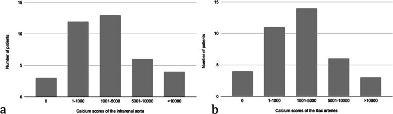
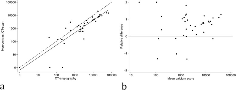
Results: Non-contrast CT revealed vascular calcification in the infrarenal aorta in 92% of KTCs and in the iliac arteries in 90% of KTCs. The calcium scores estimated from CT-angiography correlated linearly with the calcium scores based on non-contrast CT scans (infrarenal aorta: R2 = 0.71, p < 0.0001; iliac arteries: R2 = 0.71, p < 0.0001); however, the calcium scores were higher, and volumes were lower compared to the non-contrast CT scans. The median differences in calcium scores were 1517 [48 - 6138] for the infrarenal aorta, and 2361 [59 - 8644] for the iliac arteries.
Conclusion: Vascular calcification is present in the majority of KTCs. Calcification of the infrarenal aorta and iliac arteries may be assessed using CT-angiography, though higher calcium scores and lower volumes are found compared to the non-contrast CT scan.
 求助内容:
求助内容: 应助结果提醒方式:


